Friday, December 21, 2018

Regorafenib for patients with hepatocellular carcinoma who progressed on sorafenib treatment (RESOURCE): a randomized, double-blind, placebo-controlled, phase 3 trial


Summary:


Hepatocellular carcinoma (HCC) locoregional treatment includes surgical resection, transplantation, ablation and chemoembolization. For those patients who are not candidates for such treatment, medical management with the oral multikinase inhibitor sorafenib, can provide improvement in overall survival. This study aimed to evaluate the safety and efficacy of regorafenib, for patients in whom HCC has progressed during treatment with sorafenib.

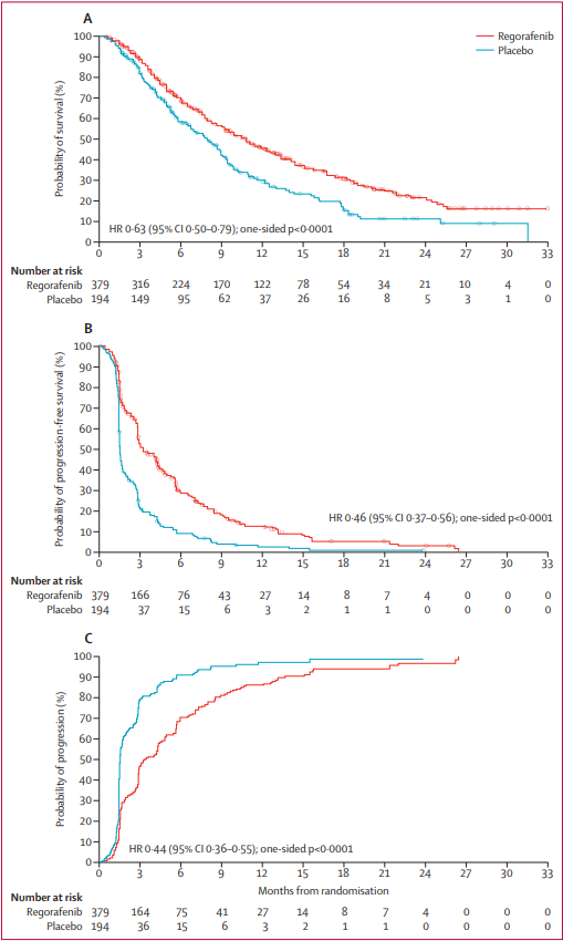
Across 152 centers and 21 countries, 573 patients with progression of HCC on sorafenib were enrolled in a randomized 2:1 regorafenib to placebo, double-blind, placebo-controlled phase 3 clinical trial. The primary endpoint was overall survival analyzed by intention to treat. Median overall survival was 10.6 months (95% CI 9.1 – 12.1) with regorafenib and 7.8 months (6.3 – 8.8) with placebo. Median progression-free survival was 3.1 months (2.8 – 4.2) with regorafenib and 1.5 months (1.4 – 1.6) with placebo by mRECIST criteria; median time to progression of 3.2 months (2.9 – 4.2) and 1.5 months (1.4 – 1.6) respectively. The safety profile of regorafenib in HCC is consistent with the use of this medication in other malignancies. The most common reason for discontinuation of regorafenib was disease progression (60%, 83% in the control group).

This study concludes that regorafenib provides a clinical improvement in overall survival in patients with progression of HCC while on sorafenib. Two patients had a complete tumor response by mRECIST criteria. This suggests that the sequential use of two multikinase inhibitors with partially overlapping target profiles can provide benefit to this patient population.


Figure 2. Kaplan-Meier analysis of overall survival (A), progression-free survival (mRECIST; B), and time to progression (mRECIST; C)

Commentary:


Regorafenib offers a viable option for patients who have failed sorafenib and has the potential to add to the current treatment armamentarium. Multiple clinical trials with second line therapies including TACE (Kudo et al., GIDEON trial, Zhang et al.), HAIC (SIRIUS trial, Terashima et al., Shao et al.) and alternative medications (Llovet et al., Zhu et al.) have demonstrated variable benefit depending on the treatment timing or drug.

The STAB Study Phase II data, recently published by Sato et al. indicates a similar class of patients (Child-Pugh class A with Barcelona Stage C disease) may benefit from concurrent TACE with sorafenib therapy with a mean overall and progression free survival of 17.3 and 5.4 months respectively, longer than those with regorafenib therapy. While this patient population was chemotherapy-naïve, it offers a potential treatment option for those patients on sorafenib prior to treatment failure. The introduction of regorafenib has the potential to change IR treatment for refractory HCC as concurrent regorafenib-TACE or alternative IR therapies may become a viable option in the future.


Bruix J, Qin S, Merle P, et al. Regorafenib for patients with hepatocellular carcinoma who progressed on sorafenib treatment (RESOURCE): a randomized, double-blind, placebo-controlled, phase 3 trial. Lancet. 2017; 389: 56-66.

Post Author:
Nicole A. Keefe, MD
Resident Physician
Department of Radiology and Medical Imaging
University of Virginia
@NikkiKeefe

Friday, December 14, 2018

Patient and Facility Demographics Related Outcomes in Early-Stage Non-Small Cell Lung Cancer Treated with Radiofrequency Ablation: A National Cancer Database Analysis 


Summary


The role of patient demographics and facility volume play with regards to survival after surgery for NSCLC has been established in multiple prior studies. However, while survival data is available for radiofrequency (RF) ablation of NSCLC, the impact of patient demographics and facility volume is not known in these patients undergoing RF ablation.

The purpose of this investigation was to evaluate the effect that these factors have on OS after RF ablation in early-stage NSCLC using the National Cancer Database (NCDB). The investigators performed a retrospective cohort study of all patients diagnosed with NSCLC from 2004 to 2014. Patients with metastatic and or nodal disease were excluded (T1aN0M0 and T1bN0M0). Additionally, patients who received chemotherapy or radiation at any time were excluded. The final cohort was a total of 967 patients. Patient demographic factors incorporated into the analysis included insurance status, income, education level and residence type. Facility demographic variables evaluated included facility type (community cancer program, comprehensive community cancer program, academic/research program, or integrated network cancer program) and facility volume. Patients were separated into those treated at high-volume centers (HVCs) and those who were not (non-HVC). After propensity score-weighted analysis of a matched cohort of 574 patients, the significant improvement in OS persisted with 1-, 3-, and 5-year OS of 89.4%, 51.2%, and 27.7%, respectively, in the HVC group, compared to 85.2%, 42.0, and 19.6%, respectively, in the non-HVC group (P=.015). Worse OS after RF ablation was significantly associated with increasing age and higher T-classification.


Commentary


This study suggests there is a significant survival benefit in patients treated with RF ablation for NSCLC in higher volume institutions. What to do with this knowledge remains the question. Additionally, more work will need to be done to look further at the specific variables accounting for this difference, apart from the higher volumes done at these centers. With all things equal, ablative technologies for NSCLC can then be pitted head to head with the current standard of care.

Click here for abstract

Lam, Alexander, et al. Patient and Facility Demographics Related Outcomes in Early-Stage Non-Small Cell Lung Cancer Treated with Radiofrequency Ablation: A National Cancer Database Analysis. Journal of Vascular and Interventional Radiology. November, 2018. Volume 29, Issue 11, 1535–1541.

Post Author: 
Zagum Bhatti, MD
Assistant Professor
Department of Radiology, Interventional Radiology Division
University of Texas Health Science Center at Houston, Houston, TX
@ZagumBhatti

Thursday, December 13, 2018

From the SIR Residents and Fellows Section (SIRRFS) Teaching Topic: Uterine Artery Embolization for the Treatment of Adenomyosis: A Systematic Review and Meta-Analysis


de Bruijn AM, Smink M, Lohle PN, Huirne JA, Twisk JW, Wong C, Schoonmade L, Hehenkamp WJ. Uterine Artery Embolization for the Treatment of Adenomyosis: A Systematic Review and Meta-Analysis. Journal of Vascular and Interventional Radiology. 2017 Oct 9.

Click here for abstract

This evidence-based review article evaluated the effect of uterine artery embolization (UAE) on symptom reduction in adenomyosis with respect to length of follow-up (< 12 months or >12 months) and whether patients had pure (adenomyosis alone) or combined (adenomyosis and fibroids) disease. The meta-analysis supported that uterine artery embolization (UAE) for both pure and combined adenomyosis resulted in significant short-term and long-term symptom improvement. There were limitations in that no randomized controlled trials were available, many included studies were retrospectively evaluated, many studies had a small sample size, and many studies offered vague description of methodology. As well, the sample size of most included studies was not sufficient to draw solid conclusions. This called for future trials to use standardized outcomes and validated questionnaires to render outcomes that can be extrapolated in systematic reviews.


Figure. Improvement of clinical symptoms (%).

Clinical Pearls


What is the pathophysiology of adenomyosis?

Adenomyosis defined as heterotopic endometrium in the myometrium and has diffuse, segmental, superficial, and deep forms. It is typically considered a disease of multiparous women in the late reproductive years. Though the etiology is still not well known, adenomyosis is thought to occur from either endomyometrial invagination of the endometrium or de novo from mullerian rests. There is also evidence that the junctional zone (JZ), which separates the subendometrial myometrium from the outer myometrium (appears as a dark band on T2WI), plays a role, as disease in the JZ predisposes to secondary infiltration of endomyometrial elements. Aside from older, multiparous women, JZ distortion can also be found in young, nulliparous women with heavy periods. A JZ > 12 mm is highly predictive of disease and < 8 mm essentially excludes it. Six studies in the meta-analysis reported a JZ reduction of 13.7%-38% after UAE. Smeets et al reported a thicker JZ at baseline to be a possible predictor of UAE failure in patients with adenomyosis. Patients who underwent hysterectomy owing to persisting symptoms compared with patients with clinical improvement showed a statistically significant thicker JZ at baseline (P = .028) and during 3 months (P = .034) of follow-up. Nijenhuis et al reported the mean JZ thickness at baseline not to be significantly thicker in groups with insufficient response (P = .17); however, it was statistically significant thicker in 4 patients needing additional therapy (P = .004).

What other gynecological pathologies are associated with adenomyosis?

Concurrent benign, estrogen-dependent gynecologic pathology, namely leiomyoma and endometriosis, is often present, increasing operative risks for major gynecologic surgery. There is also a known association between recurrent fetal loss and adenomyosis, though there is no direct evidence that adenomyosis is a risk factor for spontaneous abortion. The authors of this study state that in addition to the use of standardized outcomes and validated questionnaires, future trials should focus on fertility results in patients with symptomatic adenomyosis. Only two of the thirty-four studies in their meta-analysis reported fertility outcomes in terms of pregnancy outcome after UAE. Kim et al reported on 5 pregnancies. Two of these patients underwent elective abortion owing to unwanted pregnancies. The remaining three patients carried to term and showed no signs of uteroplacental vascular insufficiency or abnormal uterine contraction during labor or postpartum. The pregnancies resulted in 2 vaginal deliveries and 1 elective cesarean delivery because of previous cesarean delivery. The average weight of neonates was 3.2 kg (range, 3.1–3.4 kg). Yao et al described 1 pregnancy after UAE reporting termination of the pregnancy after 26 weeks. The reason for termination was not reported. Of 99 patients, 6 (6%) became pregnant after UAE. There is a paucity of high-quality data documenting the ability to conceive following UAE. Studies have reported between 48-59% live birth pregnancy rates, although the data is limited and influenced by age and current fertility status. Based on limited data, a discussion should take place with the patient about her fertility goals and a referral to a reproductive endocrinologist may also be helpful.

Why are symptoms of adenomyosis?

Though adenomyosis may be asymptomatic, it typically produces the symptoms of dysmenorrhea, dyspareunia, and chronic pelvic pain. These are also the most common symptoms of the other estrogen-dependent, benign gynecologic pathologies of endometriosis, leiomyoma, and endometrial polyp, which makes delineation of the symptoms profile difficult if the processes are concurrent. The included studies in this meta-analysis reported improvement of clinical symptoms in 83.1% (872 of 1,049) of patients. Only five of the thirty-four studies used the standardized Uterine Fibroid Symptom and Quality of Life (UFS-QOL; Spies JB, Coyne K, Guaou Guaou N, Boyle D, Skyrnarz-Murphy K, Gonzalves SM. The UFS-QOL, a new disease-specific symptom and health-related quality of life questionnaire for leiomyomata. Obstet Gynecol 2002; 99:290–300) questionnaire to evaluate health-related quality of life (HRQOL) and symptom severity. This use of this questionnaire should be used in future trials to render outcomes that can be extrapolated in systematic reviews.

Question to Consider


What are complications of UAE?

There were 20 studies that reported on complications in 615 patients. Abdominal pain occurring directly following the procedure until 2 weeks after the procedure was described in 361 of 413 patients (87.4%). Persistent amenorrhea was reported in 28 of 445 patients (6.3%) in 13 studies. All of these patients were > 40 years of age. A pseudoaneurysm was reported, which was treated with a thrombin injection. Spontaneous expulsion of leiomyomata was reported in ten patients. Endometritis was suspected in four patients. These patients were treated with broad-spectrum antibiotics. Deep venous thrombosis of the calf occurred in one patient; however, this patient did not require anticoagulation therapy. No deaths or other complications occurred.

For pain control, patient controlled analgesia with IV morphine, meperidine, hydromorphone, or fentanyl is effective. Some interventionalists prefer epidural or oral administration of narcotics. UAE patients often suffer nausea post-procedure and require either prophylactic administration or, as needed, delivery of antiemetic such as odansetron. Anti-inflammatory agents such as IV ketorolac or oral ibuprofen periprocedurally or postoperatively are commonly used. Some physicians prefer administering steroids pre-procedurally, as well, to help limit post-embolization symptoms.

The risk of infection in UAE is low with reported rates of 0.2-1% of patients. Infection usually occurs in a delayed fashion, one to two weeks after the procedure. There have been several cases of fatal sepsis occurring after UAE in patients who did not receive prophylactic antibiotics. Currently, SIR recommends prophylaxis with a single dose of 1 mg Cefazolin IV as a common choice to cover typical organisms causing infection, including skin flora and E. Coli

1. Genc M, Genc B, Cengiz H. Adenomyosis and accompanying gynecological pathologies. Arch Gynecol Obstet 2015; 291:877–881.
Brosens JJ, de Souza NM, Barker FG. Uterine junctional zone: function and disease. Lancet 1995; 346:558.

3. Spies JB, Coyne K, Guaou Guaou N, Boyle D, Skyrnarz-Murphy K, Gonzalves SM. The UFS-QOL, a new disease-specific symptom and health-related quality of life questionnaire for leiomyomata. Obstet Gynecol 2002; 99:290–300.

4. LoVerme W, James M, Ho Ahn S. “Patient Evaluation: Genitourinary System.” Patient Care in Vascular and Interventional Radiology. Third Edition. Society of Interventional Radiology, 2016. 99-111. Print.

Post Author:
Rajat Chand
Radiology Resident
John H. Stroger, Jr. Hospital of Cook County
Wednesday, December 12, 2018

Outcomes of Surgical Resection after Radioembolization for Hepatocellular Carcinoma


Summary


This is a retrospective study evaluating all patients who underwent both radioembolization (TARE) and subsequent hepatic resection in the setting of hepatocellular carcinoma (HCC). The study population (31 patients) was derived from 155 patients with HCC who underwent TARE but were also potential surgical resection candidates [preserved liver function, unilobar disease (or bilobar within Milan), no portal vein thrombus, no metastases]. The remaining patients, of the 155, were either transplanted (33) or considered for transplant (91).

The study population was made of 94% CP A and 77% BCLC Stage A disease. Technique of TARE was either lobar at a median dose of 128 Gy or segmental ablative at a medium dose of 254 Gy. 27/31 patients had a single treatment. There were no radiation-induced complications.

In addition to surgical outcomes, the authors looked extensively at the future liver remnant (FLR) following Y-90 therapy, as 25/31 patients underwent a major hepatic resection. FLR was calculated from the pre-TARE to the pre-surgical imaging. FLR increased significantly from 35% to 45% in those who received lobar Y-90 (20/25) and from 32% to 34% in those who received segmentectomy (5/25).

EASL response rates after TARE were evaluated. Stable disease was present in 39%, partial response 29%, and complete response 29%. Surgical resection after TARE occurred a median of 2.9 months after initial therapy. 24/31 patients had > 90% necrosis based on pathologic specimens.

In follow-up, 9/31 patients developed either a systemic or liver recurrence (all of whom had primary tumors > 5 cm), preferentially so in those who were EASL stable disease or had less pathologic necrosis. These findings yielded hazard ratios to develop recurrence for EASL responders of 0.18 (CI 0.5-0.7) and for 50-99% pathologic necrosis patients of 0.12 (CI 0.02-0.6). Median time to recurrence was 34 months. Median survival time was 96% at 1 and 2 years in the setting of a median follow-up time of 13 months.


Commentary


From a large number of TARE patients, this study was able to parse out a subset who underwent both TARE and subsequent surgical resection. The authors successfully demonstrate acceptable outcomes of surgical resection in the setting of prior radioembolization. They also effectively elucidate that response after Y-90 therapy by both imaging and pathologic criteria is a harbinger of future risk of recurrence after surgery.
Indirectly, this article seems to also be about the use of radioembolization to increase FLR, thus allowing surgical resection. The authors mention the potential benefits of this use, i.e. allowing local tumor control and mitigating the potential increase in local tumor growth, should one use for example portal vein embolization.
Currently TARE is often utilized in the setting non-operative HCC candidates. The authors expanded its use in this study, it seems using it as a potential bridge to resection for those in whom the FLR was insufficient to allow for surgical resection. Directly addressing this potential use of Y-90 would be helpful in future studies, as the current data only reflects the outcomes of those who successfully underwent surgery. Indeed, perhaps the success of this procedure in terms of FLR growth is much lower than theorized.

Click here for abstract

Gabr A, Abouchaleh N, Ali R, et al. Outcomes of surgical resection after radioembolization for hepatocellular carcinoma. J Vasc Interv Radiol. 2018; 29(11): 1502-1510.

Post Author:
Daniel P. Sheeran, MD
Assistant Professor
Department of Radiology and Medical Imaging
Division of Vascular and Interventional Radiology
University of Virginia

Tuesday, December 11, 2018

Diagnostic accuracy of noncontrast MR angiography protocols at 3T for the detection and characterization of lower extremity peripheral arterial disease


Summary


A clinical study evaluating non-contrast magnetic resonance angiography (MRA) to gadolinium (Gd)-based contrast MRA for lower extremity peripheral artery disease (PAD) in 20 patients was performed at 3T (vs. 1.5 T as most published diagnostic accuracy studies). Two established non-contrast MRA protocols were compared to conventional Gadolinium (Gd)-contrast enhanced MRA: (1) Quadruple inversion-recovery (QIR) Electrocardiogram combined with (ECG)-gated fast spin echo (ECG-FSE) and (2) quiescent-interval single-shot (QISS).

Imaged patients (n = 20) had intermittent claudication (Fontaine stage II) with an ankle-brachial index of < 0.9. Exclusion criteria included rest pain, nonhealing ulcers, estimated glomerular filtration rate < 30 mL/min/1.73 m2, pregnancy, claustrophobia, clinical instability or any contraindication to 3T MR imaging.

In this study, imaging datasets were reviewed in a blinded fashion by two radiologists. Non-Gd protocols demonstrated comparable diagnostic accuracy with high specificity compared to Gd-MRA protocols.


Commentary


The authors present a well-designed study evaluating forthcoming techniques in non-contrast enhanced MRA for the evaluation of lower extremity peripheral vascular disease. The techniques demonstrate information comparable to conventional contrast-enhanced MRA.

In the PAD patient population, a tool obviating the need for contrast is attractive as patients regularly have concomitant degrees of renal dysfunction. Ultimately, this study demonstrated that non-contrast MRA is a promising vascular imaging tool.

Limitations appropriately addressed by the authors include patient characteristics that may prevent or limit application of MR imaging, including body habitus, inability to follow breathing instructions, or claustrophobia. Furthermore, comparative imaging and information with non-MR focused vascular imaging tools was not performed.

Post Author:
David M. Tabriz, MD
Assistant Professor
Rush University Medical Center
@DrDaveTabriz

Tuesday, December 4, 2018

Comparison between Surgical and Endovascular Hemodialysis Arteriovenous Fistula Interventions and Associated Costs


Summary


A retrospective comparison of outcomes between patients who underwent surgical versus endovascular creation of an arteriovenous fistula (SAVF vs EAVF) was performed. Outcomes studied included rate of intervention, associated heath care cost of maintaining the fistula and the intervention free survival at 1 year after fistula creation.

The data for the surgical cohort (SAVF) was obtained from the United States Renal Data System standard analytical files(USRDS). The data for the endovascular cohort (EAVF) was obtained from the NEAT study. Medicare payment rates for any intervention performed after creation of the fistula was used as a proxy for cost comparison (both technical and professional). Two groups of patients in each cohort were described. Incident patients are not yet on dialysis but have a fistula created in anticipation where as prevalent patients are on dialysis. The two cohorts were matched 1:1 using a propensity matching system. In the incident group, 27 and in the prevalent group 33 patients from each cohort (SAVF vs EAVF) were compared.

In the incident group the event rates were 0.74/Patient Year(PY) for the EAVF cohort whereas it was 7.22/PY for the SAVF cohort(P<0.0001) resulting in a cost difference of $16,494/patient. In the prevalent group the event rates were 0.46/PY and 4.1/PY respectively for the EAVF and the SAVF cohorts (P<0.0001) resulting in a cost difference of $13,389/patient. Additional sensitivity analysis removing central venous catheter related events for the incident group brought the event rates lower for both cohorts at 0.62/PY(EAVF) and 3.84/PY(SAVF) with a cost difference of $11,290/patient. A time to event analysis at 1 year showed 70% of the incident group who underwent endovascular fistula creation were free from intervention compared to 18% in the surgical cohort. These numbers for the prevalent group were 62% and 18%. All these values were significant at P<0.05.


Commentary


Health care costs curtailment without compromising quality of care is the mantra of the future. Value of a procedure is determined by the simple formula of outcome/cost. This paper compared propensity matched patients who underwent endovascular versus surgical fistula creation for outcomes and costs. The findings favor endovascular fistula creation to be more valuable than surgical creation. This is applicable in both patient population ie those who are not on dialysis (incident group) and those who are on dialysis (prevalent group). The intervention rates and costs are much lower for the EAVF cohort irrespective of whether they are or are not on dialysis.

The authors appropriately address the limitations of this study. The NEAT study (EAVF cohort) were patients from Canada, Australia and New Zealand whereas the USRDS (SAVF cohort) are from the US. A propensity match may not capture differences in processes of care such as multidisciplinary care. The NEAT study had a large number of incident patients (almost 50%) which may skew the data in favor of EAVF as these patients are not on dialysis. The study is retrospective and the cost are not the real numbers but Medicare rates. Notwithstanding these deficiencies, the study is a powerful indicator that endovascular creation of AV fistulas is a valuable tool. Further randomized studies addressing the outcomes will be needed to bring this new tool to be the procedure of choice for creation of vascular access.

Click here for abstract

Arnold RJG, Han Y, Balakrishnan R, et al. Comparison between Surgical and Endovascular Hemodialysis Arteriovenous Fistula Interventions and Associated Costs. J Vasc Interv Radiol 2018; 29:1558-66 e2.


Post Author:
Anil K Pillai, MD
Associate Professor and Section Chief,
University of Texas Health Science Center
Anil Pillai@AnkupiMD