Monday, January 31, 2022

Stability of Retained Inferior Vena Cava Filter Fragments after Filter Removal

Stability of Retained Inferior Vena Cava Filter Fragments after Filter Removal


Clinical question
How stable are retained IVC filter fragments over time?

Take away point
98% (50/51) retained IVC filter components were stable in position by imaging at a mean imaging time of 875 days (note the single filter component deemed unstable never embolized; it rotated 180 degrees from its intramuscular position and was therefore felt to be “unstable”). The findings suggest that IVC filter fragments are predominantly stable over time.

Reference
Puller, H. F., Stavropoulos, S. W., & Trerotola, S. O. (2021). Stability of Retained Inferior Vena Cava Filter Fragments after Filter Removal. Journal of Vascular and Interventional Radiology, 32(10), 1457-1462.

Single center retrospective

Funding source
No reported funding

Setting
Hospital of the University of Pennsylvania

Figure

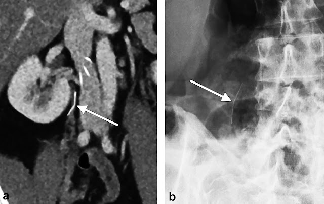
Figure 1(a) Initial computed tomography imaging of a single unstable fragment (leg) located in the right psoas muscle. The fragment concavity can be seen facing laterally (white arrow). (b) The 34-month follow-up X-ray imaging displaying interval rotation of the same fragment. The concavity can now be seen facing posteromedially (white arrow).

Summary


In 2010 there was a safety communication from the FDA due to reports of IVC filter migration and component fracture/embolization. The recommendation at the time was to consider removal of all IVC filters, if possible, once the risk of pulmonary embolism had been mitigated. The safety communication was updated in 2014 to again report the risks of IVC filter component fracture, as well as to update providers with data that IVC filter benefit was now felt to decrease at dwell times greater than 54 days.

The group of authors of the current study have previously discussed retrieval of IVC filter components in 2017. In that study, a retrospective analysis spanning 11 years. 65 patients were identified who had 116 different filter components which were fractured. The authors were able to remove 81% of the fragments that they targeted (primarily using endobronchial forceps), however, the overall removal rate of was only 54% (63/116) due to components that were not able to be targeted e.g extravascular or distal pulmonary artery. The study was useful in that it demonstrated that filter fragment retrieval could be performed safely and with high technical success. However, given that many of the filter components were left in situ, the stability of these malpositioned filter components over time remained unanswered.

The current study therefore aimed to answer that question. A retrospective review was performed at a single center from 2005 through 2020. The review identified 37 patients who presented for a complex filter removal. These individuals had fractured filter components at the time of referral which were either unable to be removed during the filter retrieval procedure, or, were not targeted for removal due to difficult or inaccessible locations. Patients were included in the review only if imaging was available at a minimum of 28 days after the last imaging of the fracture component.

In total the 37 patients had 51 total fractured filter components for review. Of the 51 components, the locations were as follows:
  • Pericaval (extravascular): 20
  • Pulmonary artery: 10
  • Wall of IVC: 5
  • Heart: 5
  • Other: 11
Of the 51 fracture filter components, 98% (50/51) were deemed stable and entirely unchanged in position at a mean imaging interval of 875 days. The authors concluded that, when asymptomatic, fractured IVC filter components are predominantly stable and can be safely followed on an intermediate- to long-term basis.

Commentary


This study informs us that IVC filter components which are fractured are very likely to be stable over time. Most of the filter components in the study seemed to be embedded. For example, these components were ones that persisted despite prior attempts at removal with endobronchial forceps or, the components were in areas that no attempt at removal was even attempted e.g. the extravascular space or distal pulmonary arteries.

These findings indicate that we can reassure patients that retained extravascular IVC filter components, or filter material that is partially intravascular but embedded (resistant to advanced removal technique, including forceps), should be followed conservatively with imaging. The expectation is that these filter components will not migrate nor will they be clinically relevant in any way.

Lastly, the importance of IVC filter tracking and retrieval programs should be highlighted given the frequency of fracture which is the reason this study was needed in the first place.

Post Author
Robert Elliott, DO
Interventional Radiology Resident, PGY6
University of Rochester Medical Center, Rochester, New York

Edited and formatted by @NingchengLi
Interventional Radiology Resident
Dotter Institute, Oregon Health and Science University

Friday, January 28, 2022

Endovascular Celiac Denervation for Glycemic Control in Patients with Type 2 Diabetes Mellitus

Endovascular Celiac Denervation for Glycemic Control in Patients with Type 2 Diabetes Mellitus


Clinical question
Can a novel method of endovascular denervation (EDN) by radiofrequency ablation of the celiac and peri-celiac aortic ganglionic sympathetic nerves improve glycemic control in patients with type II diabetes mellitus (DM2)?

Take away point
The investigators found a clinically significant improvement in glycemic control and insulin resistance in a small single-arm cohort of 11 patients with DM2 treated with endovascular denervation in the celiac artery and adjacent abdominal aorta, without any significant vascular or gastrointestinal complications. This is the first study in humans using this technology for the control and treatment of DM2.

Study design
Prospective, single-arm, non-randomized, proof-of-concept pilot study of 11 patients with DM2.

References
Pan T, Li L, Wei Q, Wang Z, Zhang Q, Qian Y, Li R, Liu DC, Wang Y, Sun ZL, Teng GJ. Endovascular Celiac Denervation for Glycemic Control in Patients with Type 2 Diabetes Mellitus. J Vasc Interv Radiol. 2021 Nov;32(11):1519-1528.e2. doi: 10.1016/j.jvir.2021.07.023. Epub 2021 Aug 6. PMID: 34364991.

Click here for abstract

Funding source
Academic research grants including:
National Major Program of Research and Development of China (2018YFA0704100)
Major Program of Scientific Instrument of the National Natural Science Foundation of China (81827805)
Clinical Medicine Center of Jiangsu Province (Innovation Section) (YXZXA2016005)
National Natural Science Foundation of Jiangsu Youth Project (BK20200365)

Setting
Academic hospital, Nanjing Drum Tower Hospital, Medical School of Nanjing University

Figures


The novel single-use denervation catheter (Golden Leaf, Shanghai, China) with 6-electrodes is illustrated. The accessory RF generator is not shown. The catheter is composed of a central wire (1), mesh tube (2), electrodes (3), mesh stent (4), and a protective sheath (5). The catheter is first exposed, then deployed, after which the electrodes are apportioned to the blood vessel wall, and EDN is then performed.


The denervation device is positioned in the celiac artery, where 1 cycle of RF ablation occurs.


The denervation device is positioned in the aorta around the celiac artery, where 2 cycles of RF ablation occur.

Summary


The study enrolled 11 patients with DM2 seeking to improve glycemic control via endovascular denervation of the celiac and peri-celiac sympathetic postganglionic nerves, which suppresses insulin secretion from pancreatic β islet cells. This treatment methodology is based on animal study that showed improved glucose tolerance in rats after celiac ganglionectomy, as well as incidental clinical results in a subset of cancer patients treated with postganglionic nerve block who developed improved glycemic control. The study enrolled patients aged 18-75 with DM2 for at least 5 years, using at least one form of diabetic medication, with the majority of patients using insulin. A 6-electrode radiofrequency endovascular catheter was used to target and ablate the post-ganglionic sympathetic nerves at the celiac and superior mesenteric ganglia. Using a transfemoral approach, an aortogram was performed to position the catheter at the level of the celiac artery and superior mesenteric artery. The EDN device was then deployed, and allowed to ablate for 3 cycles of 120 seconds each at a temperature of 60 degrees Celsius, with a maximum impedance of 400 Ω, after which a completion arteriogram was performed to ensure no vascular injury.

All of the participants achieved technical success, and there were no cases of vascular injury. Approximately 36% of patients developed immediate arterial vasospasm, which resolved shortly thereafter, while a few others developed self-limiting nausea and vomiting, abdominal distention, or constipation. Six-month follow-up demonstrated a significant decrease in the mean HbA1c by 1.9% as well as a significantly decreased homeostasis model assessment of insulin resistance (HOMA-IR) from 13.3 (IQR 5.9-46.1) to 6.0 (IQR 3.1-11.9). Mean fasting plasma glucose also decreased, and 36.4% of patients were able to discontinue at least 1 class of diabetic medication. Weight and body mass index did not undergo significant change, however there was significant decrease in alanine aminotransferase (baseline mean 31.0 IU/L to 24.0 IU/L at 6 months) and gamma glutamyl transpeptidase levels (baseline mean 47.0 IU/L to 27.0 IU/L at 6 months). These reductions suggest an additional modulating effect of EDN on nonalcoholic fatty liver disease (NAFLD), consistent with a recent animal study showing that manipulation of hepatic sympathetic innervation may be a novel therapeutic strategy for NAFLD.

Commentary


The Centers for Disease Control and Prevention estimates that nearly 1 in 10 Americans have diabetes, making it a disease with significant epidemiologic burden and many associated health complications. Thus, any novel treatment to alleviate this immense, growing public health burden could be hugely impactful. Endovascular denervation has been gaining traction in treating another prevalent disease, specifically resistant/refractory hypertension (HTN). This technique is a minimally invasive descendant of surgical sympathectomy for the same indication, which was promising but seemed overly invasive in the face of evolving pharmaceutical therapies. Thus far, results in randomized control trials of renal denervation (RDN) have been promising but mixed, some of which may be attributable to operator inexperience with the technique. Since hypertension and DM2 are frequently comorbid diseases, studies were able to assess the role of RDN in DM2 and showed early promise for achieving glycemic control. However further studies have not shown reliable success for denervation of the renal arteries.

This study by Pan et al. coopts this technique in a different anatomic location, the celiac and peri-celiac aorta, with early promising results as an endovascular approach to achieving glycemic control in patients with DM2. Given that this is a small, nonrandomized study without a control arm, the results must be considered in the context of known limitations of this study design, which the authors acknowledge. Additionally, the study measured results only up to 6 months’ post-procedure, and the authors note that nerve regeneration, which has been observed in animal models, may occur and alter long-term effects. Nonetheless, the excellent safety profile, albeit in a small non-randomized sample set, and decreased HOMA-IR, plasma glucose, liver function tests, and de-escalation in diabetic medications is promising. If reproducible results can be elicited from randomized control trials performed by experienced groups, there will be demonstrable utility for this technique either as a standalone treatment or in a combination with pharmacologic therapy for medication de-escalation. This an encouraging research space because it offers an exciting new role for Interventional Radiologists in treating diseases with a growing public health burden in a potentially single-session, minimally invasive manner.

Additional Reading:
Symplicity HTN-1 Investigators. Catheter-based renal sympathetic denervation for resistant hypertension: durability of blood pressure reduction out to 24 months. Hypertension. 2011 May;57(5):911-7. doi: 10.1161/HYPERTENSIONAHA.110.163014. Epub 2011 Mar 14. PMID: 21403086.

Post Author
Surbhi B. Trivedi, MD
Diagnostic Radiology Resident (PGY-3)
Department of Diagnostic Radiology
University of Illinois Hospital and Health Sciences System
Chicago, Illinois
Twitter: @surbhitrivedi3

Edited and formatted by @NingchengLi
Interventional Radiology Resident
Dotter Institute, Oregon Health and Science University

Monday, January 24, 2022

Thyroid Embolization for Nonsurgical Treatment of Nodular Goiter: A Single-Center Experience in 56 Consecutive Patients

Thyroid Embolization for Nonsurgical Treatment of Nodular Goiter: A Single-Center Experience in 56 Consecutive Patients


Clinical question
Is thyroid artery embolization (TAE) for the treatment of nodular goiter (NG) safe and effective?

Take away point
TAE safely and effectively reduces thyroid gland size and volume.

Reference
Yilmaz S, Habib HA, Yildiz A, Altunbas H. Thyroid Embolization for Nonsurgical Treatment of Nodular Goiter: A Single-Center Experience in 56 Consecutive Patients. J Vasc Interv Radiol. 2021; 32:1449-1456. doi.org/10.1016/j.jvir.2021.06.025

Click here for abstract

Study design
Single-institution retrospective, observational cohort study of 56 patients undergoing TAE for NG.

Funding Source
No reported funding.

Setting
Private practice center, Antalya, Turkey.

Figure

Anatomy of the thyroid arteries. The bilateral superior and inferior thyroid arteries are the main vascular supply to the thyroid gland (a). The right and left inferior thyroid arteries (white arrows, b and c) originate from the thyrocervical trunks and the left superior thyroid artery (white arrow, d) can originate from the external or common carotid arteries. The thyroid IMA artery (white arrow, e) is a small branch that may become dominant in the absence or atrophy of one of the inferior thyroid arteries.

Summary


Nodular goiter (NG) is a relatively common disorder. Given the low risk of malignant transformation, therapy is often performed for relief of mass effect symptoms or cosmetic. Minimally invasive options such as percutaneous ablation are only moderately effective in NGs >30 mL or in cases of numerous nodules or substernal extension. Therefore, surgical thyroidectomy remains the current standard therapy. Given the risk profile of thyroidectomy, thyroid artery embolization (TAE) may be a safer, less invasive alternative. The authors perform a retrospective cohort study on 56 consecutive patients undergoing TAE for NG to assess safety and efficacy.

Patients with single thyroid nodules ineligible for percutaneous laser or radiofrequency ablation and patients with multiple nodules causing compressive or cosmetic symptoms with benign FNA results (Bethesda 2-3) were included. Patients with renal insufficiency, inability to tolerate angiography, and indeterminate or atypical FNA results (Bethesda 4-6) were excluded. All patients either refused thyroidectomy or were not surgical candidates. All patients were imaged prior to treatment with either US, CT, or MRI to determine size, location, number of nodules, and intrathoracic extension. Subgroups included patients with a solitary/dominant nodule (n=20) and patients with multiple nodules (n=36).

All TAE procedures were performed via femoral arterial approach. Depending on angiography results, 2-3 branch arteries were embolized at most. In general, 2 arteries were embolized in cases of single large nodules and 3 arteries were embolized in cases of multiple nodules using 355 – 500 micrometer polyvinyl alcohol particles diluted with contrast, saline, and 5 mg papaverine. Thyroid hormone levels were periodically monitored after the procedure and follow-up imaging was completed at 6 months.

Paired t tests were used to evaluate change in total thyroid volume and thoracic extension in patients with substernal NG before and after TAE. Quality of life questionnaires were administered before and after TAE via a modified thyroid-related patient-reported outcome (Thy-PRO) survey.

A total of 56 patients met criteria with 145 of 146 targeted thyroid arteries successfully embolized. Major complications occurred in 2 patients (groin hematoma and symptomatic hyperthyroidism requiring >48-hour hospitalization) with 30-day mortality rate of 1.8% (1/56). Minor complications occurred in 25 patients.

In both subgroups, thyroid volume was significantly reduced from 80.2 mL to 25.0 mL (p<.001) in patients with a single dominant nodule, and from 147.0 mL to 62.6 mL (p<.001) in patients with multiple nodules. In patients with substernal goiter, intrathoracic extension was significantly reduced from 31.7 mm to 15.9 mm (p<.001). Overall mean ThyPRO scores improved from 155.4 to 70.4 (p<.001) at 6 months, specifically with 98% of responders (50/51) reporting that they would recommend TAE to other patients with NG.

Additionally, all patients either maintained their thyroid hormone status or improved with 19/22 patients with non-Graves hyperthyroidism able to stop their antithyroid medications. No patient developed new hypothyroidism requiring hormone replacement.

The authors discuss that, while limited in directly comparative data, their favorable outcomes support TAE as a safe and effective treatment strategy particularly in large or substernal NGs that may be technically challenging to treat via percutaneous ablation. Additionally, they discuss the superior risk profile compared to surgery with the most common minor complication being temporary hyperthyroidism likely secondary to thyroid hormone release from necrotic tissue. They compare this to the temporary hyperthyroidism often seen after radioactive iodine (RAI) therapy. But unlike RAI therapy, TAE allows for improved preservation of normal, functioning thyroid parenchyma evidenced by their cohort’s lack of postprocedural hypothyroidism necessitating hormone replacement. However, TAE remains a nonstandard therapy and various embolic techniques reported throughout the literature are compared in the discussion.

While thyroidectomy remains the standard of care, particularly in patients with NG not amenable to percutaneous ablation, TAE may be a an effective, less invasive option with a better risk profile.

Commentary


The authors evaluate the safety and effectiveness of TAE for treatment of NG via a retrospective observational cohort study. There are several limitations with this study including the small sample size and single institution design, which both limit generalizability. Additionally, the short follow up period of only 6 months likely underestimates the true complication rate and limits any conclusions drawn about long-term safety and efficacy. A longer follow up period would allow for a more robust evaluation, particularly assessing goiter recurrence and/or for need for repeat procedure. Also, the quality-of-life survey results are susceptible to selection bias as initial inclusion criteria were patients who were either not surgical candidates or who refused surgical treatment and, in that context, are more likely to report satisfaction with TAE. Lastly, the authors rightfully discuss lack of standardized procedural technique, which further limits the generalizability of the results.

Despite the limitations, the results of significantly decreased thyroid volume and intrathoracic extent are certainly remarkable for the small number complications reported, suggesting initial safety and efficacy. TAE is certainly worth further investigation as an alternative treatment to surgery for patients with NG, particularly for those with intrathoracic extension or multiple nodules that limit other minimally-invasive treatment options such as percutaneous ablation.

Post Author
Catherine (Rin) Panick, MD
Resident Physician, Integrated Interventional Radiology
Dotter Interventional Institute
Oregon Health & Science University
@MdPanick

Edited and formatted by @NingchengLi
Interventional Radiology Resident
Dotter Institute, Oregon Health and Science University

Friday, January 21, 2022

Natural History of Incidental Enhancing Nodules on Cone-Beam Computer Tomography during Transarterial Therapy of Hepatocellular carcinoma

Natural History of Incidental Enhancing Nodules on Cone-Beam Computer Tomography during Transarterial Therapy of Hepatocellular carcinoma


Clinical question
To characterize incidental enhancing nodules (IENs) seen on contrast-enhanced cone beam CT during transarterial HCC treatment.

Take away point
The majority of incidental enhancing nodules (IENs) ≥ 3 mm in patients with segmental sublobar HCC do not progress to HCC. Risk factors for progression of IENs to HCC include ≥ 4 nodules, hepatitis C, and elevated pretreatment AFP.

Reference
Elboraey M, Devcic Z, Montazeri S, Paz-Fumagalli R, McKinney J, Toskich B, et al. Natural History of Incidental Enhancing Nodules on Cone-Beam Computer Tomography during Transarterial Therapy of Hepatocellular Carcinoma. J Vasc Interv Radiol. 2021 Aug; 1(8): 1186-1192.

Click here for abstract

Study design
Single-center retrospective analysis with 100 patients with segmental sublobar HCC with contrast-enhanced CT prior to transarterial treatment.

Funding Source
No reported funding

Setting
Academic setting. Mayo Clinic Florida, Jacksonville, Florida.

Figure


Figure: Contrast enhanced MRI and CT demonstrating IENs that progressed to HCC in 6 months. (a) Contrast-enhanced magnetic resonance (MR) image demonstrated a questionable focus of enhancement in segment 5 within 30 days of cone-beam computed tomography (CT) acquisition during the transarterial treatment of hepatocellular carcinoma (HCC). (b) Contrast-enhanced cone-beam CT clearly demonstrated an incidental enhancing nodule (IEN) of ≥3 mm in segment 5. (c, d) Contrast-enhanced MR image 6 months after cone-beam CT demonstrated arterial enhancement, washout, and pseudocapsule of the progressed IEN consistent with HCC.

Summary


Hepatocellular carcinoma is diagnosed based on characteristic imaging features including arterial enhancement, delayed washout, and pseudocapsule formation on contrast-enhanced CT or MRI. Other benign and premalignant conditions such as regenerative nodules, dysplastic nodules, confluent fibrosis, and vascular shunts may also demonstrate arterial hyperenhancement, posing a diagnostic dilemma. Contrast-enhanced cone-beam CT is useful tool for detecting small hypervascular liver tumors and used during transarterial HCC therapy to identify parasitized arteries feeding the tumor. Occult enhancing lesions seen on contrast-enhanced cone beam CT may represent regenerative nodules, dysplastic nodules, or HCC and are of indeterminate significance. The authors followed incidental enhancing nodules (IENs) on contrast-enhanced CT after treatment and compared characteristics of patient with progression of IENs with those of patients without progression to HCC. Statistical analysis including the Mann-Whitney U test, chi-square test with a significance level of p <0.05, and receiver operating characteristic (ROC) curve analysis using SPSS.

One hundred-eighty patients met the study inclusion criteria of having segmental distribution sub-lobar HCC, contrast-enhanced cone-beam CT, and follow-up cross sectional imaging at least one month after the procedure. Eighty patients were excluded due to having multifocal disease, cone-beam CT not inclusive of nontarget parenchyma, lack of follow-up imaging, biphenotypic tumors, and IEN in treated territory. Of the remaining one-hundred patients, fifty-six patients had IENs on cone-beam imaging, while forty-four patients did not have IENs. One hundred fifty-four IENs were followed over a median follow-up time of two hundred eighty-two days at intervals of one, three, six, nine, and twelve months after treatment. Thirteen IENs progressed to HCC in ten patients, while one hundred forty-one IENs did not transform to HCC.

  • Patients with four or more IENs had an odds ratio (OR) of 5.9 of progression to HCC compared to patients with one to three IENs (p = 0.020).
  • Patients with increased baseline AFP had an increased risk of progression of IENs to HCC (p = 0.035, median 61.5 vs 8.6 ng/mL). Recommended cutoff of 15.5 ng/mL with AUC of 0.71, sensitivity of 80% and specificity of 67%. 
  • Patients with hepatitis C virus (HCV) infection had increased risk of progression of IENs to HCC (p = 0.015)

Characteristics of IENs that were not associated with increased risk of progression to HCC include neutrophil count (p = 0.719), lymphocyte count (p = 0.432), neutrophil-to-lymphocyte ratio (p = 0.383), and IEN size (p = 0.458).

While HCC macrovascular invasion is associated with poor prognosis, it was not associated with increased risk for IEN progression (p > 0.99). Additional characteristics of HCC that were not associated with IEN progression include HCC infiltrative patter, (p = 0.143), presence of satellites (p = 0.143), and typical HCC features (p = 0.40).

Commentary


The authors attempted to address a diagnostic dilemma encountered during the transarterial treatment of hepatocellular carcinoma: incidental enhancing nodules (IENs). The most recent guidance regarding incidental hepatic nodules is addressed in the ACR White Paper published in 2017, which recommends follow-up MRI of incidental liver masses < 1 cm in size in high-risk patients. This study provides an update to these recommendations with new data regarding the potential for IENs to progress to HCC. A prior study on IEN progression by Lucatelli et al. has reported a significantly higher rate of progression of IENs to HCC, however this is likely due to difference in study design and progression criteria. Limitations of this study include the retrospective nature, small sample size, and single institution study, which limits drawing conclusions from the data. Inherent biases of this study include a lead time bias due to a significant difference in follow-up between patients with and without progression. Further prospective studies are necessary to further evaluate the significance of incidental enhancing nodules (IENs) in the setting of hepatocellular carcinoma. The study may provide guidance in designing a prospective study to evaluate how to manage IENs in a similar patient population. Furthermore, this study suggests wider application of enhanced cone beam CT. The factors associated with increased risk of progression of IENs to HCC identified in this study (hepatitis C, IEN number >4, and elevated AFP above 15.5 ng/mL) may help guide future prospective studies and recommendations regarding this diagnostic dilemma.

Post Author
Brian Stephen Wong, MD MPH
PGY-3 Diagnostic Radiology Resident
University of Texas Medical Branch

Edited and formatted by @NingchengLi
Interventional Radiology Resident
Dotter Institute, Oregon Health and Science University

Monday, January 10, 2022

Freezing Nodal Disease: Local Control Following Percutaneous Image-Guided Cryoablation of Locoregional and Distant Lymph Node Oligiometastasis: A 10-Year, Single Center Experience

Freezing Nodal Disease: Local Control Following Percutaneous Image-Guided Cryoablation of Locoregional and Distant Lymph Node Oligiometastasis: A 10-Year, Single Center Experience


Clinical question
To retrospectively assess the technical feasibility, safety, and oncologic outcomes of percutaneous image-guided cryoablation (PCA) of locoregional and distant lymph node metastases (LNMs).

Take away point
The PCA of lymph node oligometastases is feasible and safe and offers promising local tumor control at midterm follow-up.

Reference
Autrusseau PA, Cazzato RL, Koch G, Ramamurthy N, Auloge P, De Marini P, Lipsker D, Gangi A, Garnon J. Freezing Nodal Disease: Local Control Following Percutaneous Image-Guided Cryoablation of Locoregional and Distant Lymph Node Oligometastases: A 10-Year, Single-Center Experience. J Vasc Interv Radiol. 2021 Oct;32(10):1435-1444. doi: 10.1016/j.jvir.2021.07.002. Epub 2021 Jul 13. PMID: 34271190.

Click here for abstract

Study design
Single-center retrospective descriptive analysis of patients undergoing PCA of LNMs between February 2009 and December 2019. Waiver of consent for study participation was provided following institutional review board approval. All patients gave informed consent for procedures.

Funding Source
No reported funding

Setting
Academic setting. Hôpitaux Universitaires de Strasbourg, France

Figure


Summary


Oncologic care has trended towards more aggressive treatment of metastatic disease in patients with oligometastatic and oligo-progressive lymph node metastasis (LNM). Conventional measures including surgical lymphadenectomy and radiation treatment remain mainstays but may not be feasible options for all patients due to prior surgery, challenging anatomic location of disease, morbidity, and toxicity. Percutaneous image guided cryoablation (PCA) provides a suitable option by inducing thermal injury and cell death. PCA has many advantages including excellent ablation visualization which facilitates precise procedural monitoring, synergistic combination of multiple probes to tailor ablation zone, analgesic effects, safe application following surgery or radiation therapy, and maintenance of eligibility for subsequent conventional therapy.

39 patients who underwent PCA of oligometastatic LNMs were initially identified. Exclusion criteria included subsequent cytoreductive therapy and lack of follow up. The final study population was 29 patients, 14 women and 15 men. A total of 37 procedures were performed. 17 patients had prior surgery, radiation therapy or both in the same targeted lymphatic region. 16 patients had ongoing systemic therapy at time of referral for PCA.

Procedures were completed on an inpatient basis for organizational purposes and using either a combination of US and CT guidance or US and MR guidance. All cases were monitored for intraprocedural iceball formation. Single or multiple 17-gauge probes were deployed with a minimum  5 mm ablation margin. Ancillary thermoprotective measures including hydrodissection and pneumodissection were employed when critical structures were within 10 mm of the LNM to avoid collateral thermal damage or convectional dissipation of cold energy. Cryoablation was performed using a standard double freeze protocol, 2 freezing cycles of 10 minutes with 9 minute passive thawing and 1 minute active thawing.

Imaging follow up was performed with contrast enhanced MRI at 1-, 3-, 6- and 12-months post procedure. Restaging CT and/or PET/CT were performed every 3-6 months in addition to standard oncologic follow-up including applicable tumor markers.

Important oncologic outcomes that were outlined by the study. Notable study definitions included:
  • Technical success: Complete LNM coverage by iceball, with a minimum 5 mm ablation margin on intraprocedural CT or MR imaging.
  • Primary technique efficacy: Complete LNM ablation (nonenhancement of ablation zone) at 1-month contrast enhanced MR follow-up and absence of FDG uptake during the first PET-CT follow-up (typically 6–12 weeks after the procedure.
  • Local tumor progression: New enhancement or uptake on CT, MR imaging, or PET-CT at the treated site following initial complete LNM ablation.
  • Locoregional disease progression: New locoregional LNMs (distinct from previously treated lesions) on follow-up imaging.
  • Distant disease Progression: New distant LNMs (distinct from previously treated lesions) and/or visceral metastases on follow-up imaging.
  • Local tumor progression (LTP) free survival: Time from PCA to the first radiologic evidence of LTP.
  • Disease-free survival (DFS): Time from PCA to the first radiologic evidence of disease progression at any site
  • Overall survival: Time from PCA to death from any cause.

Primary technical efficacy was 100% with complete ablation of all treated LNMs at 1 month follow up. At median follow up of 23.4 months there were 2 instances of LTP (1 patient with new regional progressive disease and 1 with isolated LTP). The 1-, 2- and 3-year LTPFS was 100%, 94.3% and 94.3% respectively. The cohort demonstrated 1-, 2- and 3-year overall survival of 96.2%, 90.5% and 70% respectively. Stratifying by location, comparing the group with locoregional LNM to the group with distant LNM revealed no statistical difference in overall survival, LTPFS or DFS at 1-, 2- or 3- years. The study also found that patients achieving remission status with local disease control were eligible for a break from systemic therapy following 54% of procedures for an average duration of 19.1 months.

Commentary


The study successfully outlines the technical feasibility and safety of PCA for LNMs with 100% technical success rate and minor complication rate of 5.4%. The noted complications were both in challenging locations with complex regional anatomy (neck and iliac/obturator LNM ablations). The results were similar to available data in recent literature with an obtained 3.7% LTP at median follow up of 23 months, compared to 4% in a smaller series. Evaluation of local disease control and recurrence rates were similar to studies focusing on HCC status post radiofrequency ablation and those evaluating stereotactic radiotherapy of LNMs. Additional direct comparison to available literature remains limited to due to heterogenous selection of response criteria between studies. Multiple factors were outlined in the study as relative strengths of PCA as a first line or salvage treatment for LNMs. The authors articulately described PCA as a useful technique in patients with high surgical morbidity, failed localization of nodal disease with alternative treatments, or patients with contraindications to full dose radiotherapy due to risks of toxicity. They also mention its utility as a mechanism to delay treatment intensification as 54% of procedures resulted in remission and halting systemic therapy. The limitations of the study include its relatively small sample size, power and retrospective design. The authors also describe selection bias, as patient selection may have leaned towards patients with better prognosis and limited disease. In the absence of a control group and due to the heterogenous stage, location and type of malignancies included, there was also a significant generalizability bias. Although the study offers promising results, future studies would be well suited to have a larger, prospective design.


Post Author
Ahmad Hashmi, MD
PGY-5 ESIR
University Hospitals Cleveland Medical Center
Case Western Reserve University
@afhashmi2

Edited and formatted by @NingchengLi
Interventional Radiology Resident
Dotter Institute, Oregon Health and Science University