Monday, July 18, 2022

Immediate and Long-Term Outcomes of Percutaneous Radiological Interventions for Hemorrhagic Complications in Acute and Chronic Pancreatitis

Immediate and Long-Term Outcomes of Percutaneous Radiological Interventions for Hemorrhagic Complications in Acute and Chronic Pancreatitis


Clinical question
What are the immediate and long-term outcomes of percutaneous radiological interventions in patients with either acute or chronic pancreatitis associated with hemorrhagic complications?

Take away point
No significant difference in outcomes—including success rates, complications, and recurrences—between patients with acute or chronic pancreatitis who received percutaneous radiological treatment for their hemorrhagic events.

Reference
Kumble Seetharama Madhusudhan, MD, FRCR, Srikanth Gopi, DM, Anand Narayan Singh, MCh, Lokesh Agarwal, MS, Deepak Gunjan, DM, Deep N. Srivastava, MD, and Pramod Kumar Garg, DM. 08/2021. Immediate and Long-Term Outcomes of Percutaneous Radiological Interventions for Hemorrhagic Complications in Acute and Chronic Pancreatitis. Journal of Vascular and Interventional Radiology. Volume 32, Issue 11, 1591-1600.

Click here for abstract

Study design
Retrospective, observational, cohort study.

Funding Source
No reported funding.

Setting
Tertiary Care Center, Department of Radiodiagnosis and Interventional Radiology, All India Institute of Medical Sciences (New Delhi, India).

Figure


Figure 1. Flowchart of the study. DSA: digital subtraction angiography, FU: follow-up.

Summary


Hemorrhagic events in cases of pancreatitis are relatively rare but can occur due to pseudoaneurysm formation, and are associated with high mortality rates. Invasive surgical intervention in such cases has high morbidity. Percutaneous embolization for patients with acute pancreatitis (AP) and chronic pancreatitis (CP) shows a high success rate but can be technically challenging. This study aimed to compare the immediate and long-term outcomes of percutaneous radiological interventions between patients with hemorrhagic complications associated with AP or CP.

The data for this retrospective cohort study was obtained from patients at a tertiary care center who received digital subtraction angiography (DSA) and endovascular intervention from January 2014 to March 2020. The mean age of patients for AP and CP were 36.5 ± 11.7 (78% [39/50] male) and 37.4 ± 11.9 (96% [54/56] male) respectively. DSA was performed via a transfemoral route. Angiography of the celiac, superior mesenteric, and inferior mesenteric arteries was performed in order to visualize the bleeding artery. The abnormal artery was then embolized using metallic coils (sandwich technique) or a mixture of glue and ethiodized oil. If endovascular embolization was unsuccessful, percutaneous embolization via ultrasound guidance was performed. Radiological interventions were correlated with clinical, angiographic, and embolization parameters. All data were analyzed using SPSS 21 software.

Technical success (complete occlusion of pseudoaneurysm or target artery) and clinical success (30-day absence of recurrent bleeding post-embolization) were not significantly different between the AP and CP groups. The overall technical success rate was 97.4% (114/117, 95% CI, 94.5-100.3%). The incidence of complications between the two groups was also not statistically different. In the AP group, 4 patients had complications post-embolization (2 splenic infarcts and 2 patients experiencing abdominal pain). In the CP group, 11 patients had complications (2 splenic infarcts, 4 splenic abscesses, and 5 patients experiencing abdominal pain). Incidence of complications was more commonly seen in older patients, female patients, bradycardic patients, and patients with a longer time-period between presentation and DSA. The higher rate of mortality in the AP group (50%, due to accompanying organ failure and sepsis) compared to the CP group (1.8%) was statistically significant (P < 0.001). The high mortality found in this study was attributed to all patients having severe presentation, with 1 or more organ failure. No significant impact was found for the embolization parameters impact on complications, recurrence of hemorrhage, and mortality. Most post-embolization patients did not experience symptoms during follow-up, and did not experience any unfavorable long-term impacts.

Commentary


This study conducted a detailed intermediate and long-term outcome comparison of interventional radiology embolization procedures in patients with hemorrhagic acute and chronic pancreatitis. Success, complications, and recurrence were comparable in the two groups. The high mortality observed in the hemorrhagic acute pancreatitis group was largely attributed to organ failure and sepsis.

Therefore, despite higher mortality rates observed in the hemorrhagic acute pancreatitis group, high technical and clinical success rates of percutaneous embolization support its use in this high-risk high-mortality population. Some questions remain to be addressed. For example, how would the comparison look like in a lower-acuity clinical setting (i.e. less severe cases or in patients without organ failure). Would a risk stratification system for acute pancreatitis help us in identifying patients who are more likely to develop hemorrhagic complications and therefore should receive aggressive fluid collection drainage. management?

Post Author
Malik Ata
Medical Student
University of Illinois College of Medicine – Chicago

Edited and formatted by @NingchengLi
Interventional Radiology Resident
Dotter Institute, Oregon Health and Science University


Friday, July 15, 2022

Prevalence of and Factors Associated with Arterial Aneurysms in Patients with Hereditary Hemorrhagic Telangiectasia: 17-Year Retrospective Series of 418 Patients

Prevalence of and Factors Associated with Arterial Aneurysms in Patients with Hereditary Hemorrhagic Telangiectasia: 17-Year Retrospective Series of 418 Patients


Clinical question
What is the prevalence of and characteristics associated with the presence of aneurysms in patients with hereditary hemorrhagic telangiectasia (HHT)?

Take away point
Compared to the general population, the prevalence of intracranial and visceral arterial aneurysms is higher in this specific cohort with HHT. Older age, unrelated AVMs and ACVRL1 mutation were associated with having an aneurysm.

Reference
Prevalence of and Factors Associated with Arterial Aneurysms in Patients with Hereditary Hemorrhagic Telangiectasia: 17-Year Retrospective Series of 418 patients (2021). Ring, N., et al. Journal of Vascular and Interventional Radiology, Volume 32: 1661-1669.

Click here for abstract

Study design
Retrospective, observational and descriptive study.

Funding Source
No funding.

Setting
Single center; HHT Center of Excellence

Figure


Summary


Hereditary Hemorrhagic Telangiectasia (HHT) is a genetic disorder characterized by vascular malformations and defined by the Curaçao criteria. Diagnosis requires inclusion of three out of four criteria: (1) recurrent epistaxis, (2) mucocutaneous telangiectasia (3) visceral lesions and (4) having a first-degree with HHT. The most well-known associated genetic mutations involve the transforming growth factor-beta (TGF-β) signaling pathway and the Endoglin (ENG) and activin A receptor type II-like 1 (ACVRL1) pathways. The TGF- β pathway has been shown to be involved in vascular remodeling and subsequent abnormal angiogenesis. Case reports suggest an increased risk of arterial aneurysms within the HHT population. This study seeks to estimate the prevalence and location of arterial aneurysms and identify any associated clinical and demographic characteristics associated with aneurysms in patients with HHT.

The study was performed at a single center, which is established as a center of excellence. A total of 418 patients with HHT were identified via chart review. The presence and characteristics (eg size, number and location) were collected. Regression modeling was performed to evaluate the following potential associations, which are known to be associated with an increased risk of aneurysms: older age, male sex, smoking, alcohol, hypertension, hyperlipidemia, genetic mutations, the presence of arteriovenous malformations unrelated to aneurysms and HHT-related genetic mutations.

Out of 418 patients, 43 (10.3%) patients had at least one aneurysm, with a total of 73 aneurysms. Eighteen (4.3%) had intracranial aneurysms and twenty patients had visceral aneurysms (4.8%), which included splenic, renal, hepatic and pancreatic aneurysms. Less than two percent were pulmonary aneurysms unrelated to AVM’s, thoracic aortic aneurysm and coronary artery aneurysms. Please see figure one for a case with multiple arterial aneurysms. Of the Curaçao criteria, only the presence of unrelated aneurysms was statistically significant. The mean Curaçao score was significantly higher in patients with aneurysms. Patients with an ACVRL1 mutation had the highest rate of arterial aneurysms (20.0%). Results from the regression analysis demonstrated presence of the ACVRL1 mutation, unrelated AVMs and older age significantly associated with having at least one aneurysm.

Meta-analyses describe the prevalence of intracranial aneurysms as 1.8-3.2% in patients without comorbidities, compared to 4.3% of patients in the present study. Prevalence of visceral artery aneurysms in the general population is 0.1-2.0%, compared to 4.8% of patients in the present study. 

A particular limitation discussed within this study surrounds the association between an unrelated AVM and an unrelated arterial aneurysm. Due to this finding, the patients underwent additional screening and may have been more likely to have an incidental aneurysm discovered. The nature of the study, single center and retrospective, limits the conclusions. The benefits of screening require further research, as the study underscores the uncertainties surrounding screening for brain AVMs in patients with HHT.

Commentary


This study demonstrates a higher rate of intracranial and visceral arterial aneurysms in their cohort of HHT patients when compared to the general population. As discussed by the authors, the largest limitation is comparing these results to the general population. The rate of aneurysms in the general population is based on research that excluded “comorbidities”. What comorbidities did they exclude and were these excluded in the present study? HHT is not an isolated diagnosis and so the small, though significant, percentage difference between the two groups could possibly be explained by comorbidities. The single-center nature of the study may also limit the conclusion regarding increased aneurysms among patients with the ACVRL1 mutation. Given HHT is an autosomal dominant genetic disorder, the same mutation may be expressed in one family living in a similar geographic area. The higher percentage of patients with ACVRL1 mutation may have allowed increased power to reach significance. 

Overall this is an excellent foundational paper for future prospective studies. Establishing an appropriate comparison group would likely strengthen the conclusions and help develop screening applications regarding frequency, age initiation, and risk stratification. 

Post Author
Marissa Stumbras, MD
Interventional Radiology Resident, PGY3
Oregon Health & Science University
@MarissaStumbras

Edited and formatted by @NingchengLi
Interventional Radiology Resident
Dotter Institute, Oregon Health and Science University


Friday, March 25, 2022

Percutaneous Arteriovenous Fistula Creation with the WavelinQ 4-French EndoAVF System: A Single-Center Retrospective Analysis of 30 Patients

Percutaneous Arteriovenous Fistula Creation with the WavelinQ 4-French EndoAVF System: A Single-Center Retrospective Analysis of 30 Patients


Clinical question
To assess the safety and efficacy of percutaneous arteriovenous fistula (pAVF) creation with WavelinQ 4F EndoAVF System.

Take away point
The WavelinQ 4F EndoAVF is a safe and effective system to create pAVFs with high maturation rate and long-term patency.

Reference
Kitrou PM, Balta L, Papachristou E, Papasotiriou M, Katsanos K, Theofanis M, Papadoulas S, Anagnostopoulos F, Georgopoulou GA, Goumenos D, Karnabatidis D. Percutaneous Arteriovenous Fistula Creation with the WavelinQ 4-French EndoAVF System: A Single-Center Retrospective Analysis of 30 Patients. J Vasc Interv Radiol. 2022 Jan;33(1):33-40. doi: 10.1016/j.jvir.2021.09.021.

Click here for abstract  

Study design
Single-center retrospective analysis with 30 patients requiring pAVFs for long-term hemodialysis.

Funding Source
The authors of this study receive consultancy fees and lecture honoraria from BD.

Setting
Academic setting. Patras University Hospital, Patras, Greece.

Figure


Figure 2a and b. Catheter alignment and different access options (only parallel approach is shown). Images showing a parallel brachial access (brachial artery and vein) of the catheters aiming to create a percutaneous arteriovenous fistula at the ulnar side (a, b). The images show the distance between the electrode (arrow) and the ceramic backstop before (a) and after (b) activation. The distance between the catheters in image (a) reflects the walls of the vessels and the tissue between the vessels. Following activation and anastomosis creation, the electrode is in contact with the ceramic backstop. In the image (a), the square orientation of the indicators at the front tip of the catheters is shown (arrowhead). 

Figure 3. Final angiogram of a procedure performed with brachial arterial access. Contrast was injected from the brachial artery. The different parts of the newly formed vascular access circuit are pointed out. 1, brachial artery; 2, radial artery; 3, anastomosis; 4, radial vein (with some spasm); 5, perforator vein; 6, cephalic vein; and 7, basilic vein. The ulnar artery (A), main ulnar artery (B), and interosseous artery (C) outside the vascular access circuit are also apparent.

Figure 4. Kaplan-Meier survival curve of the 26 cases that matured and achieved cannulation with subjects at risk.

Summary


Percutaneous arteriovenous fistulas (pAVFs) are a new endovascular technique to create hemodialysis access for patients with chronic kidney failure. Two systems are currently available: the WavelinQ EndoAVF and the Ellipsys Vascular Access System. The WavelinQ creates an anastomosis between the radial and ulnar vessels, while the Ellipsys System uses the perforating vein and proximal radial artery. pAVFs offer an additional technique for long-term hemodialysis access and may have similar outcomes with surgical arteriovenous fistula, however limited data has been published about the safety and efficacy.

Thirty patients met the inclusion criteria of being >18 years old, Stage V CKD, Target A/V diameters >2.5mm with perforating forearm vein of >2mm. Patient with active hypercoagulable state, infection, extensive vascular calcification, and contrast allergy were excluded from the study The study was a single-center, single-arm retrospective study of arteriovenous fistulas created by the 4Fr WavelinQ system.

  • 100% (30/30) patients had technical success in pAVF creation.
  • In thirty patients that underwent pAVF creation, the cannulation rate was 87% at mid-term follow-up time (mean f/u time 547 +/- 315 days).
  • 16 reinterventions (i.e. coil embolization or angioplasty) were necessary to facilitate cannulation without need for open surgical procedures.
  • 7 maintenance procedures were performed in 6 of 27 matured pAVFs.
  • 22 out of 33 (73%) pAVFs were non-thrombosed and cannulated at the end of the study.
  • Relatively safe with adverse event rate (i.e. brachial artery pseudoaneurysm) in 2/30 patients (6.7%).

Commentary


This study is one of the first to address the safety and efficacy of pAVFs. One distinct advantage of pAVFs is that the procedure does not compromise the creation of future surgical AVFs at the same site. While this technique is not risk-free, it appears that it is a safe alternative for providing hemodialysis access to patients with CKD. Other studies with the WavelinQ EndoAVF system have validated the high success rate and efficacy: 
  • Endovascular Access System Enhancements study (Berland et al. 2019; 4Fr) was the first to evaluate the current 4Fr system and reported 100% technical success in 32 patients with both ulnar and radial AVFs.
  • The FLEX study (Rajan et al. 2015; 6Fr) enrolled 33 pts with a success rate of 97% and one adverse effect (brachial pseudoaneurysm). 
  • The Novel Endovascular Access Trial (Lok et al. 2017; 6Fr) with 60 patients demonstrated a high success rate of 98% with only 2% adverse effects in ulnar pAVFs.
  • Zemela et al. 2021 (6Fr) with 35 patients reported technical success rate of 100% and one pseudoaneurysm.
  • Radosa et al. 2017 (6Fr) with 8 patients reported technical success rate of 100%.

A recent study by Inston et al. compared pAVRs (with both generations of the WavelinQ system) and surgical radiocephalic AVFs and reported comparable patency rates. A study by Shahverdyan demonstrated that WavelinQ and Ellipsys are no different in outcome. A Multicenter European/Canadian discovered that coil embolization at index reduced further need to treat. 

Similar to other studies on novel techniques of percutaneous arteriovenous fistula creation, this study's clinical value remains uncertain and practice adoption of the studied devices/techniques difficult given the small sample size, selection bias, inconsistent follow-up duration, procedural and fistula metrics incompleteness, and lack of prospective control arm(s). The authors rightfully described their findings as early experience, limited in scope, and will require external larger prospective clinical trials for validation.

Post Author
Brian Stephen Wong, MD MPH
PGY-3 Diagnostic Radiology Resident
University of Texas Medical Branch

Edited and formatted by @NingchengLi
Interventional Radiology Resident
Dotter Institute, Oregon Health and Science University

Friday, March 18, 2022

Thrombotic Risk Associated with Inferior Vena Cava Filter Placement in Patients with Heparin-Induced Thrombocytopenia

Thrombotic Risk Associated with Inferior Vena Cava Filter Placement in Patients with Heparin-Induced Thrombocytopenia


Clinical question
Are mortality and thrombotic risk increased in patients with heparin-induced thrombocytopenia (HIT) undergoing IVC filter (IVCF) placement compared to patients with either HIT or IVCF alone?

Take away point
IVC filter placement did not significantly increase risk of thromboembolism or death in patients with HIT, suggesting IVCF placement may be a viable treatment option for patients with elevated thromboembolic risk secondary to HIT in whom anticoagulation has failed or is contraindicated.

Reference
Moghbel M, Chen Z, Liu C, Rajan S, Vempaty H, and Wang S. Thrombotic Risk Associated with Inferior Vena Cava Filter Placement in Patients with Heparin-Induced Thrombocytopenia. J Vasc Interv Radiol. 2021; 32:1629-1634. doi.org/10.1016/j.jvir.2021.08.025

Click here for abstract

Study design
Retrospective, single-network cohort study of 26 patients with HIT and IVCF placement vs controls with either HIT or IVCF alone.

Funding Source
No reported funding.

Setting
Private hospital network, Kaiser Permanente Northern California, USA.

Figure

No significant difference in mortality was observed in the HIT+IVCF group versus controls.

Summary


Heparin-induced thrombocytopenia (HIT) is an adverse reaction to heparin resulting in reduced platelet count. Thromboembolic events occur in up to half of patients with HIT. Currently, it is not recommended to routinely place IVC filters (IVCFs) in patients with HIT in whom anticoagulation has failed or is contraindicated due to the paucity of outcomes data in this relatively small population. The authors perform a retrospective cohort study comparing outcomes in patients with HIT receiving IVCFs compared to patients with HIT who did not receive an IVCF and patients receiving an IVCF without a diagnosis of HIT.

Electronic medical records were queried for patients with HIT and/or IVCFs using diagnostic and procedural codes. Patient charts coded with HIT were reviewed manually and excluded if they had been miscoded. The two control groups were unable to be manually reviewed due to the immensity of the sample sizes. Patients in the HIT+IVCF group were stratified into subgroups based on likelihood of HIT (high likelihood = positive serotonin release assay or HIT antibody level >1; intermediate likelihood = HIT antibody level <1) as well as timing of IVCF placement (within 14 days of HIT diagnosis versus >14 days of HIT diagnosis). This 14-day cutoff was chosen based on estimated peak timing of HIT-associated thromboembolic risk.

Primary outcomes were 6- and 12-month mortality and thromboembolic risk, which included new or extended DVT/PE, IVC thrombosis, lower extremity phlegmasia cerulea dolens, and critical limb ischemia. Propensity score matching was performed to control for potential confounders based on demographic data. Comparisons were made using chi-squared and Fisher exact tests.

A total of 4774 patients were analyzed, 26 with HIT+IVCF, 814 with HIT without IVCF , and 3,934 with IVCF without HIT. The most common indication for IVCF placement in the HIT+IVCF group was proximal DVT/PE with contraindication to anticoagulation (n=18, 69.2%).

Mortality rates in the HIT+IVCF group were 26.9% (n=7) at 6 months and 30.8% (n=8) at 12 months compared to 29.5% (n=240) and 34.9% (n=284) in the HIT only group and 33.1% (n=1,288) and 38.2 (n=1,486) in the IVCF only group. No significant difference was noted among groups at either 6 or 12 months (p=0.11 and 0.17, respectively).

Additionally, HIT+IVCF subgroup analyses showed no significant difference in 6- or 12-month mortality between HIT likelihood (p=0.59 and 0.27, respectively) or timing of IVCF placement (p=0.69 and 0.84, respectively). Thromboembolic risk was not significantly different within these two subgroups.

The authors discuss that current guidelines recommend against the routine placement of IVCFs in patients with HIT despite limited comparative data assessing patients with HIT and IVCFs versus either alone. The results of this study suggest that patients with HIT in whom IVCFs are placed are at no higher mortality risk compared to controls nor are they at higher thromboembolic risk compared to previously reported data on patients with either HIT or IVCFs alone.

Commentary


This study compares mortality rates in patients with HIT receiving IVCFs compared to patients with either HIT or IVCF alone as well as evaluates the thromboembolic risk in patients with HIT receiving IVCFs. The authors note that current guidelines are largely based on data describing high rates of thromboembolic events in patients with HIT in whom central venous catheters or IVCFs were placed, but remark that there is limited data assessing the true thromboembolic risk of patients with HIT and IVCF compared to controls. To that end, this study aimed to more accurately characterize the thromboembolic risk and mortality in this population.

The extremely large sample sizes of the control populations, while advantageous for powering the study, did not allow for manual chart review to ensure that diagnostic and procedural codes were accurately assigned in the electronic medical records. In the HIT+IVCF group alone, 9 out of the original 39 cases (23%) were excluded due to miscoding, which raises concern regarding the accuracy of the HIT and IVCF only groups. Additionally, since the control groups were so considerably large, it was not possible for the authors to review all charts to establish thromboembolic risk in each control group. They rightfully discuss that their thromboembolic risk comparison is therefore restricted to previously published data, rather than having an internal comparison. This introduces potential biases between institutional practice patterns, patient populations, and variability over time.

Although generally quite thorough in the evaluation of the HIT+IVCF group, the authors do not mention retrieval rates of IVCFs in either this population or their controls. Given that IVCFs are known to be associated with greater thrombotic risk if not removed, it would have been interesting to see if timing of IVCF removal had any influence on thromboembolic risk or mortality.

Furthermore, although 6- and 12-month follow up periods are relatively long term in relation to duration of thromboembolic risk associated with HIT (which peaks over 14 days), it may also be of benefit to assess longer (3- or 5-year) mortality rates to validate the conclusions over an extended time period. Lastly, the retrospective design has inherent limitations where a prospective study or randomized controlled trial would offer more accurate comparison, although likely not feasible in this relatively small patient population.

The major strength of this study is its large sample size of patients with HIT receiving IVCFs compared to other studies. Their thorough review of laboratory and clinical data to determine likelihood of HIT diagnosis and timing of IVCF placement adds to the comprehensiveness of the morbidity and mortality evaluation. The scope of their discussion also appropriately addresses the limitations of the data and highlights the need for additional research and external validation.

Overall, this study sheds new light on the question of whether IVCF placement should be considered in patients with HIT in which anticoagulation has failed or is contraindicated. For patients with HIT and IVCFs, there was no significant difference in thromboembolic risk compared to rates from other studies nor in overall mortality compared to internal controls. These findings challenge current guidelines that recommend against routine placement of IVCFs in patients with HIT; however, outside validation of these results is essential prior to implementing changes in clinical practice.

Post Author
Catherine (Rin) Panick, MD
Resident Physician, Integrated Interventional Radiology
Dotter Interventional Institute
Oregon Health & Science University
@MdPanick

Edited and formatted by @NingchengLi
Interventional Radiology Resident
Dotter Institute, Oregon Health and Science University


Friday, March 11, 2022

Absolute Ethanol Embolization of Lip Arteriovenous Malformations: Observational Results from 10 Years of Experience

Absolute Ethanol Embolization of Lip Arteriovenous Malformations: Observational Results from 10 Years of Experience


Clinical question
What is the safety and efficacy of lip arteriovenous malformation (AVMs) ethanol embolization via direct puncture or transarterial catheterization technique, with or without follow-up surgical resection?

Take away point
Lip AVM treatment remains variable. Ethanol embolization of lip AVMs in single or multiple sessions was able to achieve complete devascularization in 67% of the patients and complete cosmetic resolution in 53% of patients. When including preoperative embolization followed by surgical resection, 71% achieved complete cosmetic relief and all patients endorsed major to complete improvement of functional impairment.

Reference
Su LX, Li XY, Zhao ZJ, Shao YH, Fan XD, Wen MZ, Yang XT. Absolute Ethanol Embolization of Lip Arteriovenous Malformations: Observational Results from 10 Years of Experience. J Vasc Interv Radiol. 2022 Jan;33(1):42-48.e4. doi: 10.1016/j.jvir.2021.09.004. Epub 2021 Sep 20. PMID: 34547475.

Click here for abstract

Study design
10-year retrospective, observational study of 76 patients

Funding source
(1) Fundamental research program funding of Ninth People’s Hospital affiliated to Shanghai Jiao Tong University School of Medicine (No. JYZZ076)
(2) Clinical Research Program of Ninth People’s Hospital, Shanghai Jiao Tong University School of Medicine (No. JYLJ201801 and JYLJ201911)
(3) China Postdoctoral Science Foundation (No. 2017M611585)
(4) National Natural Science Foundation of China (No. 81871458)

Setting
Urban academic center at the Shanghai Ninth People’s Hospital which is affiliated with the Shanghai Jiao Tong University School of Medicine

Figures


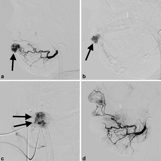
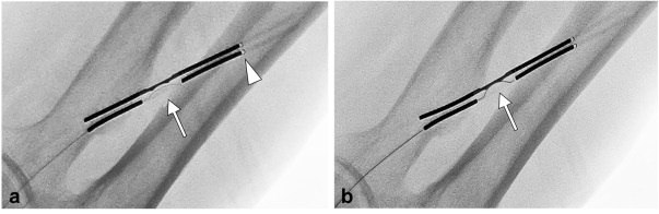
Figure 1: Intraprocedural digital subtraction angiography (DSA) of a 17-year-old male treated for a pulsating AVM. (a) A focal AVM nidus is opacified with contrast (arrow). (b) Lateral view demonstrates the 1.7 Fr microcatheter at the AVM nidus via superselective embolization from the facial artery. (c) Subsequently, direct 21-guage needle puncture was used to perform an additional embolization of the nidus. (d) The post-procedure lateral view DSA demonstrates 100% AVM devascularization.


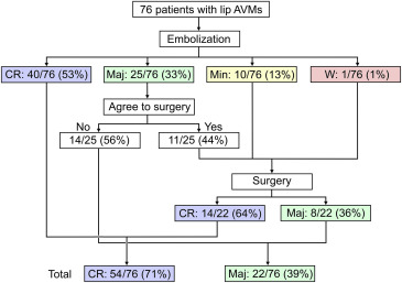
Figure 2: Of the 76 patients treated with embolization, most with multiple sessions, the majority went on to have complete cosmetic resolution either solely with ethanol embolization or after ethanol embolization and subsequent surgical resection, and 100% of patients endorses complete or major resolution of cosmetic appearance.

Summary


Lip arteriovenous malformations (AVM) may be present at birth and can enlarge over time secondary to trauma or hormonal changes. Radical surgical resection can result in functional/ cosmetic defects and may risk severe intraoperative hemorrhage. Incomplete resection fails to provide symptom relief. Thus, AVM embolization is increasingly becoming the treatment of choice.

This study utilized ethanol as a permanent embolic agent. Other studies of peripheral AVM embolization have used ethylene vinyl alcohol copolymer and n-butyl cyanoacrylate (n-BCA) glue, however reports show nidus recanalization can occur, and subcutaneous accumulation of the embolic glue can cause infection. Ethanol injection permanently destroys the vascular endothelium by causing protein denaturation and nidus thrombosis. Downsides of ethanol embolization include risks of systemic ethanol toxicity which can include cardiopulmonary arrest and disseminated intravascular coagulation (DIC).

This 10-year retrospective study includes patients with lip AVM Schobinger stage II or higher (growing or symptomatic AVM). Many patients had prior treatments (including surgical resection, sclerotherapy, propranolol, radiofrequency ablation and artery ligation), but only those at least 3 months post prior therapy were included. Other exclusion criteria included contrast or ethanol allergy and major cardiac, respiratory, renal or hepatic impairment.

Lip AVM embolization was performed under general anesthesia, with a Foley catheter in place to monitor for hemoglobinuria secondary to ethanol administration and arterial line for blood pressure monitoring. Pulmonary artery pressure was measured in those expected to have high ethanol volume administered.

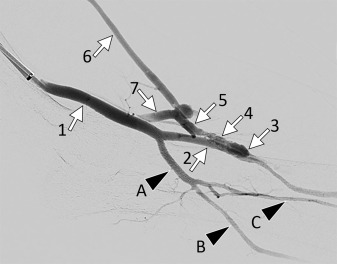
Digital subtraction angiography (DSA) was performed via superselective angiography of the external and internal carotid arteries using a 4 or 5 Fr catheter to identify the AVM nidus based on feeding vessels, draining veins, and internal blood flow. Then, a 1.7 Fr microcatheter was advanced into the nidus. If complete nidus access was not feasible by this technique alone, they performed ultrasound-guided direct puncture using a 21-guage butterfly needle. If a draining outflow vein (DOV) was identified, it was coil-embolized prior to ethanol embolization.

Required ethanol volume was estimated based on the amount of contrast injected to fill the AVM nidus. Usually 100% ethanol was used except in patients with a diffuse infiltrating AVM where diluted ethanol was used to reduce risks of systemic toxicity. In all cases, less than 1 cc/kg of ethanol was used during each session, and staged embolization was used in patients to reduce risks of ethanol toxicity. Additionally, manual pressure was applied around the target lesion to reduce non-target embolization.

The 76 patients enrolled underwent a mean of 2.3 procedures (range of 1-7 procedures) and were followed for a mean of about 2.5 years. Patients were asked to self-report cosmetic changes on a scale of 1-5 after treatment, and functional results were evaluated by oral maxillofacial surgeons.

Roughly 67% of patients had 100% AVM devascularization on arteriography. One third of patients had 76-99% devascularization and 26.3% experienced 50-75% devascularization. Symptomatically, 53% of patients had complete cosmetic improvement after embolization alone. A third of patients self-reported major cosmetic improvement, and 44% of them underwent surgery to see further improvement. Those who rated their cosmetic changes as minorly improved or worsened underwent surgery, all of whom felt they had major improvement or complete improvement postoperatively. In total, 71% had complete cosmetic resolution of their AVM and 39% had major cosmetic improvement with embolization alone or followed up by surgical resection.

Immediate post-procedure swelling was seen in almost all patients and resolved within 2 weeks. The most common complication was local skin bullae, which occurred in 9.2% of procedures and spontaneously resolved. There were 3 cases of skin necrosis, one of whom developed serious lip necrosis causing cosmetic deformity that required surgery. Two patients developed transient hemoglobinuria which was treated with IV infusion. No severe complications were reported.

Commentary


The study is limited by its retrospective nature, potential for patient selection bias, relatively small sample size and nonstandardized treatment algorithm which included a mix of embolization, coil embolization of the draining vein when present, and postprocedural surgery. A randomized control trial with a placebo arm as well as comparative groups randomly assigned to surgical or embolization therapy would be useful. Additionally, comparing the efficacy and complications of other embolic materials and combination of coils or plugs would be advantageous for clinical decision-making. Although ethanol is highly effective, rare reports of systemic effects causing DIC and cardiopulmonary arrest may be a considerable drawback for centers that do not perform lip AVM commonly.

Future studies could consider enrolling treatment-naïve patients. In this study, more than half of the patients had prior laser treatment, sclerotherapy, radiofrequency ablation or arterial embolization, performed at least 3 months prior. This may confound the results of ethanol embolization, especially in cases of prior surgical resection or arterial ligation, which can complicate future embolization access. Additionally, the study on average followed patients for 2.5 years, and though ethanol embolization is permanent when injected, AVM recurrence may occur in untreated regions of a diffuse, infiltrating AVM beyond that time frame. Another limitation is that post-procedure cosmetic results were quantified based on a subjective patient-derived scale; however, this appears to have been used in prior studies.

In conclusion, the study authors convey that lip AVM treatment can be challenging due to the nature of the AVM vascular anatomy, however multifactorial treatment options can produce acceptable results in most patients. Treatment options were customized throughout the course of therapy and tailored to patient preferences regarding cosmetic improvement. These options include multiple courses of ethanol embolization via superselective angiography and/or direct puncture technique, post-procedure surgical resection, and draining outflow vein coil embolization. The study reports that most patients had complete or major improvement of cosmetic and functional symptoms and embolization and/or surgical resection. However, a subset of patients was not able to achieve complete resolution of their cosmetic defects despite multiple sessions of embolization and surgical resection, underscoring the complicated treatment course of peripheral AVMs and the importance of accurately conveying to patients the realities of AVM treatment.

Post author
Surbhi B. Trivedi, MD
Diagnostic Radiology PGY-3 Resident
University of Illinois, Chicago
@surbhitrivedi3

Edited and formatted by @NingchengLi
Interventional Radiology Resident
Dotter Institute, Oregon Health and Science University


Friday, March 4, 2022

Clinical Safety and Efficacy of Locoregional Therapy Combined with Adoptive Transfer of Allogeneic γδ T Cells for Advanced Hepatocellular Carcinoma and Intrahepatic Cholangiocarcinoma

Clinical Safety and Efficacy of Locoregional Therapy Combined with Adoptive Transfer of Allogeneic γδ T Cells for Advanced Hepatocellular Carcinoma and Intrahepatic Cholangiocarcinoma


Clinical question
Is locoregional therapy plus adoptive transfer of allogeneic gamma delta (γδ) T cells safe and effective in treating patients with hepatocellular carcinoma (HCC) and intrahepatic cholangiocarcinoma (ICC)?

Take-away point
The combination of both locoregional ablation and adoptive transfer of allogeneic γδ cells is both safe and effective in patients with HCC and ICC.

Reference
Zhang T, Chen J, Niu L, et al. Clinical safety and efficacy of locoregional therapy combined with adoptive transfer of allogeneic γδ t cells for advanced hepatocellular carcinoma and intrahepatic cholangiocarcinoma. Journal of Vascular and Interventional Radiology. 2022;33(1):19-27.e3.

Click here for abstract

Study design
Randomized Controlled Trial

Funding source
This study was funded by grants from the National Key R&D Program of China (2018YFA0108304), National Natural Science Foundation of China (81771721, 81971505), and Fujian Provincial Health Education Joint Research Project (WKJ2016-2-20).

Setting
Single institution, Fuda Cancer Hospital of Jinan University, Guangzhou, China


Figure 5. Survival curves. Overall survival curves of hepatocellular carcinoma (HCC) patients (a). Distant progression-free survival (PFS) curves of HCC patients (b). Local PFS curves of HCC patients (ablation sites) (c). Overall survival curves of intrahepatic cholangiocarcinoma (ICC) patients (d). Distant PFS curves of ICC patients (e). Local PFS curves of ICC patients (ablation sites) (f).

Summary


Adoptive transfer of T cells has emerged as a novel treatment of malignant neoplasms. A subset of T cells known as γδ T cells has been specifically utilized to recognize and eliminate tumor cells based on tumor-specific antigens. In an allogeneic adoptive transfer strategy, the γδ T cells are provided by a donor, amplified in vitro, and subsequently infused into a patient with a known cancer (as supposed to in vivo activation by zoledronate and IL-2). Few clinical trials have been conducted in patient populations with advanced disease due to the larger tumor burden and associated immunosuppressive environment. It is postulated by the authors that the combination of locoregional ablative therapy in concert with systemic treatment using allogeneic γδ T cells may be an effective treatment. 

Cryoablation and irreversible electroporation (IRE) are locoregional therapies that have proven efficacy and safety, while also preserving tumor cell surface architecture to potentially allow immune system recognition and subsequent response. Thus, the merit behind combining these modalities with systemic immune therapy is evident. Additionally, adoptive transfer has been shown to be safe and effective in several late-stage cancers as a monotherapy as well as in combination with IRE, and infused γδ T cells are known to preferentially migrate to the liver. Thus, a study of the safety and efficacy of a combination of locoregional therapy and adoptive transfer in the treatment of hepatocellular carcinoma (HCC) and intrahepatic cholangiocarcinoma (ICC) was devised.

In this randomized controlled trial, patients between the age of 18-75 years of age with stage III or IV HCC or ICC by 7th AJCC TNM guidelines were included. The study also required patients have multifocal disease that made them unsuitable for surgery or chemotherapy. They could not have tumor invasion of the portal vein, hepatic vein trunk, or secondary branches, and had to have a projected survival of > 3 months. Additional inclusion criteria included Eastern Cooperative Oncology Group (ECOG) performance status (PS) score ≤ 2, normal laboratory parameters, and adequate hepatic and renal function. Patients with coagulation disorders, anemia, severe coronary artery disease, respiratory disease, myelosuppression, acute or chronic infection, or autoimmune disease were excluded. Patients were randomized to either the combined treatment or local treatment groups using a computer-generated random number table. Patients in the combined treatment group underwent either IRE or cryoablation followed by adoptive transfer of allogeneic γδ T cells. Patients in the local treatment group only underwent either IRE or cryoablation. IRE was used for tumors adjacent to large vessels such as the hepatic artery, portal vein, or bile ducts in the liver hilum. Other tumors were treated with cryoablation. Additionally, transarterial chemoembolization was done prior to ablation on tumors > 5 cm in size to reduce tumor size prior to locoregional therapy. In patients undergoing adoptive transfer, donor γδ cells were collected, isolated, and expanded as previously described in the literature on day 1 of treatment. The patients then underwent locoregional therapy on day 9 before receiving donated cells in 3 equal infusions, with one each on days 13, 14, and 15. This was followed by a 2nd donation on day 16 and infusions on days 28, 29, and 30. Additionally, peripheral blood was drawn on day 8 and a month after day 30 to determine CTC levels. Follow-ups included CT or MRI at 1 month and every 3 months after treatment. These were compared to imaging collected 1 week prior to locoregional therapy. Modified Response Evaluation Criteria in Solid Tumors (mRECIST) was used to evaluate treatment response. Technical success was defined as complete ablation while local progression was defined as reappearance of arterial phase enhancement of the ablated area after initial technical success. Distant progression was defined as the appearance of new intrahepatic or extrahepatic tumors or an increase of over 20% in diameter of any unablated tumors. Overall survival was defined as time between locoregional therapy and death. Additional primary endpoints included safety (adverse events), distant progression-free survival (PFS), and local PFS. Secondary endpoints were CTC levels, AFP and CA19-9 (for HCC and ICC respectively), and change in ECOG PS.

Median survival was improved in the HCC group undergoing combination therapy compared to locoregional therapy, with median OS being 13 months for combination and 8 months for locoregional therapy, 1-year OS rates of 57.3% and 7.3% respectively, and 2-year OS rates of 26.7% and 7.3% respectively (p = 0.029). Median distant PFS in combination and locoregional were 8 and 4 months, with 1-year distant PFS rates of 0% and 21.8%, respectively (p = 0.040). Differences in local PFS rates were not significant (p = 0.949, 28.6% in combination group and 14.5% in locoregional group).

For patients with ICC, differences in OS were not significant; median OS was 9 months for combination and 8 months for locoregional therapy, 1-year OS rates of 33.3% and 44.3% respectively and 2-year OS rates of 25% and 8.3% respectively (p = 0.546). Median distant PFS in combination and locoregional groups were 8 and 4 months, with 1-year distant PFS rates of 14.1% and 0%, respectively (p = 0.021). Similar to HCC groups, differences in local PFS rates were not significant (p = 0.394, 16% in combination group and 27.5% in locoregional group). The authors mention that 28.6% of ICC patients and an unspecified number HCC in their respective combination groups did not receive locoregional therapy and dropped out of the study due to progression or withdrawal of consent, prompting a protocol analysis. The results of the protocol analysis were reported as consistent with the intention-to-treat (ITT) analysis.

AFP decreased in all groups, with a more pronounced drop in the combined treatment groups (HCC: p = 0.046, ICC: p = 0.028). Change in CA-19-9 was not significantly different between the experimental and control groups. CTCs decreased in all groups, but was more pronounced in the combined treatment groups (HCC: p = 0.035, ICC: p = 0.003). ECOG PS improvements were also statistically significant in both combined treatment groups (HCC: p = 0.039, ICC: p = 0.007), whereas the locoregional groups did not significantly change after treatment.

No severe adverse events occurred during this study. Only grade 1 and 2 events were reported, with most being grade 1 (least significant). These included fever, nausea, thrombocytopenia, arrhythmia, hemothorax, pneumothorax, pleural effusion, and fatigue. The majority of these were grade 1 and all adverse events were resolved spontaneously or with symptomatic treatment. Most events were associated with locoregional therapy, and there was no significant difference in incidence between the two groups. There was no significant difference in lab markers for hepatic or renal function.

Commentary


This study aimed to demonstrate safety and explore hypothesized efficacy of adoptive transfer of healthy volunteers' (allogeneic) γδ T cells in combination with locoregional therapies in the treatment of advanced HCC and ICC. This was done by employing previously-described techniques and timelines that have been proven safe and modestly effective. Results of this study demonstrated statistically significant if modest improvements in distant PFS for both HCC and ICC patients, but no differences were seen in local PFS. Additionally, OS was improved after combination therapy for HCC patients, but not for ICC patients. It is supposed that the lack of difference in local PFS is due to the primary contribution of locoregional therapy to this metric in all groups. Despite this, PS did improve in both combination groups while not in locoregional groups. It is hard to say, however, if this is clinically significant. The authors also posited that the significantly pronounced decrease in CTCs in both combination groups was likely secondary to systemic effects of γδ T cells and also likely the reason for the differences in distant PFS. Indeed, decrease in CTCs is a previously-demonstrated effect of γδ T cell infusion, and is known to be associated with increased PFS as well as impaired metastasis and recurrence. The decrease in AFP in HCC patients with combination therapy and the respective lack of decrease in CA19-9 in ICC patients, in concert with the less-pronounced survival differences in ICC patients, further suggests that combination therapy may be less effective in treating ICC. ICC is also acknowledged as a morbid diagnosis that progresses rapidly, is refractory to first-line treatments, and is often diagnosed in advanced stages, and thus may require a larger sample to determine true efficacy. 

While this trial was an RCT, it does have some limitations. One is that a higher proportion of patients in the combination groups did not undergo intended therapy. Although the PP and ITT analyses were consistent, this still may affect true therapeutic effect. Additionally, the study was open-label without allocation-concealment, which undermines the randomization design. Finally, the use of both IRE or cryoablation, with their different utilities and mechanisms of action, may have resulted in unknown effects on the data. Future trials will benefit from larger sample size, standardization of locoregional therapy or subgroup analysis of the modalities used, and double-blinding to ensure randomization.

Post Author
Jared Edwards, MD
General Medical Officer
Medical Readiness Division
Naval Surface Forces Pacific, San Diego, CA
@JaredRayEdwards

Edited and formatted by @NingchengLi
Interventional Radiology Resident
Dotter Institute, Oregon Health and Science University

Monday, January 31, 2022

Stability of Retained Inferior Vena Cava Filter Fragments after Filter Removal

Stability of Retained Inferior Vena Cava Filter Fragments after Filter Removal


Clinical question
How stable are retained IVC filter fragments over time?

Take away point
98% (50/51) retained IVC filter components were stable in position by imaging at a mean imaging time of 875 days (note the single filter component deemed unstable never embolized; it rotated 180 degrees from its intramuscular position and was therefore felt to be “unstable”). The findings suggest that IVC filter fragments are predominantly stable over time.

Reference
Puller, H. F., Stavropoulos, S. W., & Trerotola, S. O. (2021). Stability of Retained Inferior Vena Cava Filter Fragments after Filter Removal. Journal of Vascular and Interventional Radiology, 32(10), 1457-1462.

Single center retrospective

Funding source
No reported funding

Setting
Hospital of the University of Pennsylvania

Figure

Figure 1(a) Initial computed tomography imaging of a single unstable fragment (leg) located in the right psoas muscle. The fragment concavity can be seen facing laterally (white arrow). (b) The 34-month follow-up X-ray imaging displaying interval rotation of the same fragment. The concavity can now be seen facing posteromedially (white arrow).

Summary


In 2010 there was a safety communication from the FDA due to reports of IVC filter migration and component fracture/embolization. The recommendation at the time was to consider removal of all IVC filters, if possible, once the risk of pulmonary embolism had been mitigated. The safety communication was updated in 2014 to again report the risks of IVC filter component fracture, as well as to update providers with data that IVC filter benefit was now felt to decrease at dwell times greater than 54 days.

The group of authors of the current study have previously discussed retrieval of IVC filter components in 2017. In that study, a retrospective analysis spanning 11 years. 65 patients were identified who had 116 different filter components which were fractured. The authors were able to remove 81% of the fragments that they targeted (primarily using endobronchial forceps), however, the overall removal rate of was only 54% (63/116) due to components that were not able to be targeted e.g extravascular or distal pulmonary artery. The study was useful in that it demonstrated that filter fragment retrieval could be performed safely and with high technical success. However, given that many of the filter components were left in situ, the stability of these malpositioned filter components over time remained unanswered.

The current study therefore aimed to answer that question. A retrospective review was performed at a single center from 2005 through 2020. The review identified 37 patients who presented for a complex filter removal. These individuals had fractured filter components at the time of referral which were either unable to be removed during the filter retrieval procedure, or, were not targeted for removal due to difficult or inaccessible locations. Patients were included in the review only if imaging was available at a minimum of 28 days after the last imaging of the fracture component.

In total the 37 patients had 51 total fractured filter components for review. Of the 51 components, the locations were as follows:
  • Pericaval (extravascular): 20
  • Pulmonary artery: 10
  • Wall of IVC: 5
  • Heart: 5
  • Other: 11
Of the 51 fracture filter components, 98% (50/51) were deemed stable and entirely unchanged in position at a mean imaging interval of 875 days. The authors concluded that, when asymptomatic, fractured IVC filter components are predominantly stable and can be safely followed on an intermediate- to long-term basis.

Commentary


This study informs us that IVC filter components which are fractured are very likely to be stable over time. Most of the filter components in the study seemed to be embedded. For example, these components were ones that persisted despite prior attempts at removal with endobronchial forceps or, the components were in areas that no attempt at removal was even attempted e.g. the extravascular space or distal pulmonary arteries.

These findings indicate that we can reassure patients that retained extravascular IVC filter components, or filter material that is partially intravascular but embedded (resistant to advanced removal technique, including forceps), should be followed conservatively with imaging. The expectation is that these filter components will not migrate nor will they be clinically relevant in any way.

Lastly, the importance of IVC filter tracking and retrieval programs should be highlighted given the frequency of fracture which is the reason this study was needed in the first place.

Post Author
Robert Elliott, DO
Interventional Radiology Resident, PGY6
University of Rochester Medical Center, Rochester, New York

Edited and formatted by @NingchengLi
Interventional Radiology Resident
Dotter Institute, Oregon Health and Science University

Friday, January 28, 2022

Endovascular Celiac Denervation for Glycemic Control in Patients with Type 2 Diabetes Mellitus

Endovascular Celiac Denervation for Glycemic Control in Patients with Type 2 Diabetes Mellitus


Clinical question
Can a novel method of endovascular denervation (EDN) by radiofrequency ablation of the celiac and peri-celiac aortic ganglionic sympathetic nerves improve glycemic control in patients with type II diabetes mellitus (DM2)?

Take away point
The investigators found a clinically significant improvement in glycemic control and insulin resistance in a small single-arm cohort of 11 patients with DM2 treated with endovascular denervation in the celiac artery and adjacent abdominal aorta, without any significant vascular or gastrointestinal complications. This is the first study in humans using this technology for the control and treatment of DM2.

Study design
Prospective, single-arm, non-randomized, proof-of-concept pilot study of 11 patients with DM2.

References
Pan T, Li L, Wei Q, Wang Z, Zhang Q, Qian Y, Li R, Liu DC, Wang Y, Sun ZL, Teng GJ. Endovascular Celiac Denervation for Glycemic Control in Patients with Type 2 Diabetes Mellitus. J Vasc Interv Radiol. 2021 Nov;32(11):1519-1528.e2. doi: 10.1016/j.jvir.2021.07.023. Epub 2021 Aug 6. PMID: 34364991.

Click here for abstract

Funding source
Academic research grants including:
National Major Program of Research and Development of China (2018YFA0704100)
Major Program of Scientific Instrument of the National Natural Science Foundation of China (81827805)
Clinical Medicine Center of Jiangsu Province (Innovation Section) (YXZXA2016005)
National Natural Science Foundation of Jiangsu Youth Project (BK20200365)

Setting
Academic hospital, Nanjing Drum Tower Hospital, Medical School of Nanjing University

Figures


The novel single-use denervation catheter (Golden Leaf, Shanghai, China) with 6-electrodes is illustrated. The accessory RF generator is not shown. The catheter is composed of a central wire (1), mesh tube (2), electrodes (3), mesh stent (4), and a protective sheath (5). The catheter is first exposed, then deployed, after which the electrodes are apportioned to the blood vessel wall, and EDN is then performed.


The denervation device is positioned in the celiac artery, where 1 cycle of RF ablation occurs.


The denervation device is positioned in the aorta around the celiac artery, where 2 cycles of RF ablation occur.

Summary


The study enrolled 11 patients with DM2 seeking to improve glycemic control via endovascular denervation of the celiac and peri-celiac sympathetic postganglionic nerves, which suppresses insulin secretion from pancreatic β islet cells. This treatment methodology is based on animal study that showed improved glucose tolerance in rats after celiac ganglionectomy, as well as incidental clinical results in a subset of cancer patients treated with postganglionic nerve block who developed improved glycemic control. The study enrolled patients aged 18-75 with DM2 for at least 5 years, using at least one form of diabetic medication, with the majority of patients using insulin. A 6-electrode radiofrequency endovascular catheter was used to target and ablate the post-ganglionic sympathetic nerves at the celiac and superior mesenteric ganglia. Using a transfemoral approach, an aortogram was performed to position the catheter at the level of the celiac artery and superior mesenteric artery. The EDN device was then deployed, and allowed to ablate for 3 cycles of 120 seconds each at a temperature of 60 degrees Celsius, with a maximum impedance of 400 Ω, after which a completion arteriogram was performed to ensure no vascular injury.

All of the participants achieved technical success, and there were no cases of vascular injury. Approximately 36% of patients developed immediate arterial vasospasm, which resolved shortly thereafter, while a few others developed self-limiting nausea and vomiting, abdominal distention, or constipation. Six-month follow-up demonstrated a significant decrease in the mean HbA1c by 1.9% as well as a significantly decreased homeostasis model assessment of insulin resistance (HOMA-IR) from 13.3 (IQR 5.9-46.1) to 6.0 (IQR 3.1-11.9). Mean fasting plasma glucose also decreased, and 36.4% of patients were able to discontinue at least 1 class of diabetic medication. Weight and body mass index did not undergo significant change, however there was significant decrease in alanine aminotransferase (baseline mean 31.0 IU/L to 24.0 IU/L at 6 months) and gamma glutamyl transpeptidase levels (baseline mean 47.0 IU/L to 27.0 IU/L at 6 months). These reductions suggest an additional modulating effect of EDN on nonalcoholic fatty liver disease (NAFLD), consistent with a recent animal study showing that manipulation of hepatic sympathetic innervation may be a novel therapeutic strategy for NAFLD.

Commentary


The Centers for Disease Control and Prevention estimates that nearly 1 in 10 Americans have diabetes, making it a disease with significant epidemiologic burden and many associated health complications. Thus, any novel treatment to alleviate this immense, growing public health burden could be hugely impactful. Endovascular denervation has been gaining traction in treating another prevalent disease, specifically resistant/refractory hypertension (HTN). This technique is a minimally invasive descendant of surgical sympathectomy for the same indication, which was promising but seemed overly invasive in the face of evolving pharmaceutical therapies. Thus far, results in randomized control trials of renal denervation (RDN) have been promising but mixed, some of which may be attributable to operator inexperience with the technique. Since hypertension and DM2 are frequently comorbid diseases, studies were able to assess the role of RDN in DM2 and showed early promise for achieving glycemic control. However further studies have not shown reliable success for denervation of the renal arteries.

This study by Pan et al. coopts this technique in a different anatomic location, the celiac and peri-celiac aorta, with early promising results as an endovascular approach to achieving glycemic control in patients with DM2. Given that this is a small, nonrandomized study without a control arm, the results must be considered in the context of known limitations of this study design, which the authors acknowledge. Additionally, the study measured results only up to 6 months’ post-procedure, and the authors note that nerve regeneration, which has been observed in animal models, may occur and alter long-term effects. Nonetheless, the excellent safety profile, albeit in a small non-randomized sample set, and decreased HOMA-IR, plasma glucose, liver function tests, and de-escalation in diabetic medications is promising. If reproducible results can be elicited from randomized control trials performed by experienced groups, there will be demonstrable utility for this technique either as a standalone treatment or in a combination with pharmacologic therapy for medication de-escalation. This an encouraging research space because it offers an exciting new role for Interventional Radiologists in treating diseases with a growing public health burden in a potentially single-session, minimally invasive manner.

Additional Reading:
Symplicity HTN-1 Investigators. Catheter-based renal sympathetic denervation for resistant hypertension: durability of blood pressure reduction out to 24 months. Hypertension. 2011 May;57(5):911-7. doi: 10.1161/HYPERTENSIONAHA.110.163014. Epub 2011 Mar 14. PMID: 21403086.

Post Author
Surbhi B. Trivedi, MD
Diagnostic Radiology Resident (PGY-3)
Department of Diagnostic Radiology
University of Illinois Hospital and Health Sciences System
Chicago, Illinois
Twitter: @surbhitrivedi3

Edited and formatted by @NingchengLi
Interventional Radiology Resident
Dotter Institute, Oregon Health and Science University

Monday, January 24, 2022

Thyroid Embolization for Nonsurgical Treatment of Nodular Goiter: A Single-Center Experience in 56 Consecutive Patients

Thyroid Embolization for Nonsurgical Treatment of Nodular Goiter: A Single-Center Experience in 56 Consecutive Patients


Clinical question
Is thyroid artery embolization (TAE) for the treatment of nodular goiter (NG) safe and effective?

Take away point
TAE safely and effectively reduces thyroid gland size and volume.

Reference
Yilmaz S, Habib HA, Yildiz A, Altunbas H. Thyroid Embolization for Nonsurgical Treatment of Nodular Goiter: A Single-Center Experience in 56 Consecutive Patients. J Vasc Interv Radiol. 2021; 32:1449-1456. doi.org/10.1016/j.jvir.2021.06.025

Click here for abstract

Study design
Single-institution retrospective, observational cohort study of 56 patients undergoing TAE for NG.

Funding Source
No reported funding.

Setting
Private practice center, Antalya, Turkey.

Figure

Anatomy of the thyroid arteries. The bilateral superior and inferior thyroid arteries are the main vascular supply to the thyroid gland (a). The right and left inferior thyroid arteries (white arrows, b and c) originate from the thyrocervical trunks and the left superior thyroid artery (white arrow, d) can originate from the external or common carotid arteries. The thyroid IMA artery (white arrow, e) is a small branch that may become dominant in the absence or atrophy of one of the inferior thyroid arteries.

Summary


Nodular goiter (NG) is a relatively common disorder. Given the low risk of malignant transformation, therapy is often performed for relief of mass effect symptoms or cosmetic. Minimally invasive options such as percutaneous ablation are only moderately effective in NGs >30 mL or in cases of numerous nodules or substernal extension. Therefore, surgical thyroidectomy remains the current standard therapy. Given the risk profile of thyroidectomy, thyroid artery embolization (TAE) may be a safer, less invasive alternative. The authors perform a retrospective cohort study on 56 consecutive patients undergoing TAE for NG to assess safety and efficacy.

Patients with single thyroid nodules ineligible for percutaneous laser or radiofrequency ablation and patients with multiple nodules causing compressive or cosmetic symptoms with benign FNA results (Bethesda 2-3) were included. Patients with renal insufficiency, inability to tolerate angiography, and indeterminate or atypical FNA results (Bethesda 4-6) were excluded. All patients either refused thyroidectomy or were not surgical candidates. All patients were imaged prior to treatment with either US, CT, or MRI to determine size, location, number of nodules, and intrathoracic extension. Subgroups included patients with a solitary/dominant nodule (n=20) and patients with multiple nodules (n=36).

All TAE procedures were performed via femoral arterial approach. Depending on angiography results, 2-3 branch arteries were embolized at most. In general, 2 arteries were embolized in cases of single large nodules and 3 arteries were embolized in cases of multiple nodules using 355 – 500 micrometer polyvinyl alcohol particles diluted with contrast, saline, and 5 mg papaverine. Thyroid hormone levels were periodically monitored after the procedure and follow-up imaging was completed at 6 months.

Paired t tests were used to evaluate change in total thyroid volume and thoracic extension in patients with substernal NG before and after TAE. Quality of life questionnaires were administered before and after TAE via a modified thyroid-related patient-reported outcome (Thy-PRO) survey.

A total of 56 patients met criteria with 145 of 146 targeted thyroid arteries successfully embolized. Major complications occurred in 2 patients (groin hematoma and symptomatic hyperthyroidism requiring >48-hour hospitalization) with 30-day mortality rate of 1.8% (1/56). Minor complications occurred in 25 patients.

In both subgroups, thyroid volume was significantly reduced from 80.2 mL to 25.0 mL (p<.001) in patients with a single dominant nodule, and from 147.0 mL to 62.6 mL (p<.001) in patients with multiple nodules. In patients with substernal goiter, intrathoracic extension was significantly reduced from 31.7 mm to 15.9 mm (p<.001). Overall mean ThyPRO scores improved from 155.4 to 70.4 (p<.001) at 6 months, specifically with 98% of responders (50/51) reporting that they would recommend TAE to other patients with NG.

Additionally, all patients either maintained their thyroid hormone status or improved with 19/22 patients with non-Graves hyperthyroidism able to stop their antithyroid medications. No patient developed new hypothyroidism requiring hormone replacement.

The authors discuss that, while limited in directly comparative data, their favorable outcomes support TAE as a safe and effective treatment strategy particularly in large or substernal NGs that may be technically challenging to treat via percutaneous ablation. Additionally, they discuss the superior risk profile compared to surgery with the most common minor complication being temporary hyperthyroidism likely secondary to thyroid hormone release from necrotic tissue. They compare this to the temporary hyperthyroidism often seen after radioactive iodine (RAI) therapy. But unlike RAI therapy, TAE allows for improved preservation of normal, functioning thyroid parenchyma evidenced by their cohort’s lack of postprocedural hypothyroidism necessitating hormone replacement. However, TAE remains a nonstandard therapy and various embolic techniques reported throughout the literature are compared in the discussion.

While thyroidectomy remains the standard of care, particularly in patients with NG not amenable to percutaneous ablation, TAE may be a an effective, less invasive option with a better risk profile.

Commentary


The authors evaluate the safety and effectiveness of TAE for treatment of NG via a retrospective observational cohort study. There are several limitations with this study including the small sample size and single institution design, which both limit generalizability. Additionally, the short follow up period of only 6 months likely underestimates the true complication rate and limits any conclusions drawn about long-term safety and efficacy. A longer follow up period would allow for a more robust evaluation, particularly assessing goiter recurrence and/or for need for repeat procedure. Also, the quality-of-life survey results are susceptible to selection bias as initial inclusion criteria were patients who were either not surgical candidates or who refused surgical treatment and, in that context, are more likely to report satisfaction with TAE. Lastly, the authors rightfully discuss lack of standardized procedural technique, which further limits the generalizability of the results.

Despite the limitations, the results of significantly decreased thyroid volume and intrathoracic extent are certainly remarkable for the small number complications reported, suggesting initial safety and efficacy. TAE is certainly worth further investigation as an alternative treatment to surgery for patients with NG, particularly for those with intrathoracic extension or multiple nodules that limit other minimally-invasive treatment options such as percutaneous ablation.

Post Author
Catherine (Rin) Panick, MD
Resident Physician, Integrated Interventional Radiology
Dotter Interventional Institute
Oregon Health & Science University
@MdPanick

Edited and formatted by @NingchengLi
Interventional Radiology Resident
Dotter Institute, Oregon Health and Science University

Friday, January 21, 2022

Natural History of Incidental Enhancing Nodules on Cone-Beam Computer Tomography during Transarterial Therapy of Hepatocellular carcinoma

Natural History of Incidental Enhancing Nodules on Cone-Beam Computer Tomography during Transarterial Therapy of Hepatocellular carcinoma


Clinical question
To characterize incidental enhancing nodules (IENs) seen on contrast-enhanced cone beam CT during transarterial HCC treatment.

Take away point
The majority of incidental enhancing nodules (IENs) ≥ 3 mm in patients with segmental sublobar HCC do not progress to HCC. Risk factors for progression of IENs to HCC include ≥ 4 nodules, hepatitis C, and elevated pretreatment AFP.

Reference
Elboraey M, Devcic Z, Montazeri S, Paz-Fumagalli R, McKinney J, Toskich B, et al. Natural History of Incidental Enhancing Nodules on Cone-Beam Computer Tomography during Transarterial Therapy of Hepatocellular Carcinoma. J Vasc Interv Radiol. 2021 Aug; 1(8): 1186-1192.

Click here for abstract

Study design
Single-center retrospective analysis with 100 patients with segmental sublobar HCC with contrast-enhanced CT prior to transarterial treatment.

Funding Source
No reported funding

Setting
Academic setting. Mayo Clinic Florida, Jacksonville, Florida.

Figure


Figure: Contrast enhanced MRI and CT demonstrating IENs that progressed to HCC in 6 months. (a) Contrast-enhanced magnetic resonance (MR) image demonstrated a questionable focus of enhancement in segment 5 within 30 days of cone-beam computed tomography (CT) acquisition during the transarterial treatment of hepatocellular carcinoma (HCC). (b) Contrast-enhanced cone-beam CT clearly demonstrated an incidental enhancing nodule (IEN) of ≥3 mm in segment 5. (c, d) Contrast-enhanced MR image 6 months after cone-beam CT demonstrated arterial enhancement, washout, and pseudocapsule of the progressed IEN consistent with HCC.

Summary


Hepatocellular carcinoma is diagnosed based on characteristic imaging features including arterial enhancement, delayed washout, and pseudocapsule formation on contrast-enhanced CT or MRI. Other benign and premalignant conditions such as regenerative nodules, dysplastic nodules, confluent fibrosis, and vascular shunts may also demonstrate arterial hyperenhancement, posing a diagnostic dilemma. Contrast-enhanced cone-beam CT is useful tool for detecting small hypervascular liver tumors and used during transarterial HCC therapy to identify parasitized arteries feeding the tumor. Occult enhancing lesions seen on contrast-enhanced cone beam CT may represent regenerative nodules, dysplastic nodules, or HCC and are of indeterminate significance. The authors followed incidental enhancing nodules (IENs) on contrast-enhanced CT after treatment and compared characteristics of patient with progression of IENs with those of patients without progression to HCC. Statistical analysis including the Mann-Whitney U test, chi-square test with a significance level of p <0.05, and receiver operating characteristic (ROC) curve analysis using SPSS.

One hundred-eighty patients met the study inclusion criteria of having segmental distribution sub-lobar HCC, contrast-enhanced cone-beam CT, and follow-up cross sectional imaging at least one month after the procedure. Eighty patients were excluded due to having multifocal disease, cone-beam CT not inclusive of nontarget parenchyma, lack of follow-up imaging, biphenotypic tumors, and IEN in treated territory. Of the remaining one-hundred patients, fifty-six patients had IENs on cone-beam imaging, while forty-four patients did not have IENs. One hundred fifty-four IENs were followed over a median follow-up time of two hundred eighty-two days at intervals of one, three, six, nine, and twelve months after treatment. Thirteen IENs progressed to HCC in ten patients, while one hundred forty-one IENs did not transform to HCC.

  • Patients with four or more IENs had an odds ratio (OR) of 5.9 of progression to HCC compared to patients with one to three IENs (p = 0.020).
  • Patients with increased baseline AFP had an increased risk of progression of IENs to HCC (p = 0.035, median 61.5 vs 8.6 ng/mL). Recommended cutoff of 15.5 ng/mL with AUC of 0.71, sensitivity of 80% and specificity of 67%. 
  • Patients with hepatitis C virus (HCV) infection had increased risk of progression of IENs to HCC (p = 0.015)

Characteristics of IENs that were not associated with increased risk of progression to HCC include neutrophil count (p = 0.719), lymphocyte count (p = 0.432), neutrophil-to-lymphocyte ratio (p = 0.383), and IEN size (p = 0.458).

While HCC macrovascular invasion is associated with poor prognosis, it was not associated with increased risk for IEN progression (p > 0.99). Additional characteristics of HCC that were not associated with IEN progression include HCC infiltrative patter, (p = 0.143), presence of satellites (p = 0.143), and typical HCC features (p = 0.40).

Commentary


The authors attempted to address a diagnostic dilemma encountered during the transarterial treatment of hepatocellular carcinoma: incidental enhancing nodules (IENs). The most recent guidance regarding incidental hepatic nodules is addressed in the ACR White Paper published in 2017, which recommends follow-up MRI of incidental liver masses < 1 cm in size in high-risk patients. This study provides an update to these recommendations with new data regarding the potential for IENs to progress to HCC. A prior study on IEN progression by Lucatelli et al. has reported a significantly higher rate of progression of IENs to HCC, however this is likely due to difference in study design and progression criteria. Limitations of this study include the retrospective nature, small sample size, and single institution study, which limits drawing conclusions from the data. Inherent biases of this study include a lead time bias due to a significant difference in follow-up between patients with and without progression. Further prospective studies are necessary to further evaluate the significance of incidental enhancing nodules (IENs) in the setting of hepatocellular carcinoma. The study may provide guidance in designing a prospective study to evaluate how to manage IENs in a similar patient population. Furthermore, this study suggests wider application of enhanced cone beam CT. The factors associated with increased risk of progression of IENs to HCC identified in this study (hepatitis C, IEN number >4, and elevated AFP above 15.5 ng/mL) may help guide future prospective studies and recommendations regarding this diagnostic dilemma.

Post Author
Brian Stephen Wong, MD MPH
PGY-3 Diagnostic Radiology Resident
University of Texas Medical Branch

Edited and formatted by @NingchengLi
Interventional Radiology Resident
Dotter Institute, Oregon Health and Science University

Monday, January 10, 2022

Freezing Nodal Disease: Local Control Following Percutaneous Image-Guided Cryoablation of Locoregional and Distant Lymph Node Oligiometastasis: A 10-Year, Single Center Experience

Freezing Nodal Disease: Local Control Following Percutaneous Image-Guided Cryoablation of Locoregional and Distant Lymph Node Oligiometastasis: A 10-Year, Single Center Experience


Clinical question
To retrospectively assess the technical feasibility, safety, and oncologic outcomes of percutaneous image-guided cryoablation (PCA) of locoregional and distant lymph node metastases (LNMs).

Take away point
The PCA of lymph node oligometastases is feasible and safe and offers promising local tumor control at midterm follow-up.

Reference
Autrusseau PA, Cazzato RL, Koch G, Ramamurthy N, Auloge P, De Marini P, Lipsker D, Gangi A, Garnon J. Freezing Nodal Disease: Local Control Following Percutaneous Image-Guided Cryoablation of Locoregional and Distant Lymph Node Oligometastases: A 10-Year, Single-Center Experience. J Vasc Interv Radiol. 2021 Oct;32(10):1435-1444. doi: 10.1016/j.jvir.2021.07.002. Epub 2021 Jul 13. PMID: 34271190.

Click here for abstract

Study design
Single-center retrospective descriptive analysis of patients undergoing PCA of LNMs between February 2009 and December 2019. Waiver of consent for study participation was provided following institutional review board approval. All patients gave informed consent for procedures.

Funding Source
No reported funding

Setting
Academic setting. Hôpitaux Universitaires de Strasbourg, France

Figure


Summary


Oncologic care has trended towards more aggressive treatment of metastatic disease in patients with oligometastatic and oligo-progressive lymph node metastasis (LNM). Conventional measures including surgical lymphadenectomy and radiation treatment remain mainstays but may not be feasible options for all patients due to prior surgery, challenging anatomic location of disease, morbidity, and toxicity. Percutaneous image guided cryoablation (PCA) provides a suitable option by inducing thermal injury and cell death. PCA has many advantages including excellent ablation visualization which facilitates precise procedural monitoring, synergistic combination of multiple probes to tailor ablation zone, analgesic effects, safe application following surgery or radiation therapy, and maintenance of eligibility for subsequent conventional therapy.

39 patients who underwent PCA of oligometastatic LNMs were initially identified. Exclusion criteria included subsequent cytoreductive therapy and lack of follow up. The final study population was 29 patients, 14 women and 15 men. A total of 37 procedures were performed. 17 patients had prior surgery, radiation therapy or both in the same targeted lymphatic region. 16 patients had ongoing systemic therapy at time of referral for PCA.

Procedures were completed on an inpatient basis for organizational purposes and using either a combination of US and CT guidance or US and MR guidance. All cases were monitored for intraprocedural iceball formation. Single or multiple 17-gauge probes were deployed with a minimum  5 mm ablation margin. Ancillary thermoprotective measures including hydrodissection and pneumodissection were employed when critical structures were within 10 mm of the LNM to avoid collateral thermal damage or convectional dissipation of cold energy. Cryoablation was performed using a standard double freeze protocol, 2 freezing cycles of 10 minutes with 9 minute passive thawing and 1 minute active thawing.

Imaging follow up was performed with contrast enhanced MRI at 1-, 3-, 6- and 12-months post procedure. Restaging CT and/or PET/CT were performed every 3-6 months in addition to standard oncologic follow-up including applicable tumor markers.

Important oncologic outcomes that were outlined by the study. Notable study definitions included:
  • Technical success: Complete LNM coverage by iceball, with a minimum 5 mm ablation margin on intraprocedural CT or MR imaging.
  • Primary technique efficacy: Complete LNM ablation (nonenhancement of ablation zone) at 1-month contrast enhanced MR follow-up and absence of FDG uptake during the first PET-CT follow-up (typically 6–12 weeks after the procedure.
  • Local tumor progression: New enhancement or uptake on CT, MR imaging, or PET-CT at the treated site following initial complete LNM ablation.
  • Locoregional disease progression: New locoregional LNMs (distinct from previously treated lesions) on follow-up imaging.
  • Distant disease Progression: New distant LNMs (distinct from previously treated lesions) and/or visceral metastases on follow-up imaging.
  • Local tumor progression (LTP) free survival: Time from PCA to the first radiologic evidence of LTP.
  • Disease-free survival (DFS): Time from PCA to the first radiologic evidence of disease progression at any site
  • Overall survival: Time from PCA to death from any cause.

Primary technical efficacy was 100% with complete ablation of all treated LNMs at 1 month follow up. At median follow up of 23.4 months there were 2 instances of LTP (1 patient with new regional progressive disease and 1 with isolated LTP). The 1-, 2- and 3-year LTPFS was 100%, 94.3% and 94.3% respectively. The cohort demonstrated 1-, 2- and 3-year overall survival of 96.2%, 90.5% and 70% respectively. Stratifying by location, comparing the group with locoregional LNM to the group with distant LNM revealed no statistical difference in overall survival, LTPFS or DFS at 1-, 2- or 3- years. The study also found that patients achieving remission status with local disease control were eligible for a break from systemic therapy following 54% of procedures for an average duration of 19.1 months.

Commentary


The study successfully outlines the technical feasibility and safety of PCA for LNMs with 100% technical success rate and minor complication rate of 5.4%. The noted complications were both in challenging locations with complex regional anatomy (neck and iliac/obturator LNM ablations). The results were similar to available data in recent literature with an obtained 3.7% LTP at median follow up of 23 months, compared to 4% in a smaller series. Evaluation of local disease control and recurrence rates were similar to studies focusing on HCC status post radiofrequency ablation and those evaluating stereotactic radiotherapy of LNMs. Additional direct comparison to available literature remains limited to due to heterogenous selection of response criteria between studies. Multiple factors were outlined in the study as relative strengths of PCA as a first line or salvage treatment for LNMs. The authors articulately described PCA as a useful technique in patients with high surgical morbidity, failed localization of nodal disease with alternative treatments, or patients with contraindications to full dose radiotherapy due to risks of toxicity. They also mention its utility as a mechanism to delay treatment intensification as 54% of procedures resulted in remission and halting systemic therapy. The limitations of the study include its relatively small sample size, power and retrospective design. The authors also describe selection bias, as patient selection may have leaned towards patients with better prognosis and limited disease. In the absence of a control group and due to the heterogenous stage, location and type of malignancies included, there was also a significant generalizability bias. Although the study offers promising results, future studies would be well suited to have a larger, prospective design.


Post Author
Ahmad Hashmi, MD
PGY-5 ESIR
University Hospitals Cleveland Medical Center
Case Western Reserve University
@afhashmi2

Edited and formatted by @NingchengLi
Interventional Radiology Resident
Dotter Institute, Oregon Health and Science University