Minimally Invasive Image-Guided Ablation, Osteoplasty, Reinforcement, and Internal Fixation (AORIF) for Osteolytic Lesions in the Pelvis and Periarticular Regions of Weight-Bearing Bones
Is AORIF a safe and effective surgical alternative to treat lytic metastases in periarticular load-bearing bones?
Take-away point
AORIF is a safe and effective treatment for symptomatic osteolytic metastases in load-bearing bones.
Reference
Lee FY, Latich I, Toombs C, et al. Minimally Invasive Image-Guided Ablation, Osteoplasty, Reinforcement, and Internal Fixation (AORIF) for Osteolytic Lesions in the Pelvis and Periarticular Regions of Weight-Bearing Bones. J Vasc Interv Radiol. 2020;31(4):649–658.e1. doi:10.1016/j.jvir.2019.11.029
Click here for abstract
Study design
Single-center, prospective clinical cohort
Funding source
Research support from the National Cancer Institute and National Institute of Arthritis and Musculoskeletal and Skin Diseases
Setting
Academic hospital, Yale School of Medicine, United States.
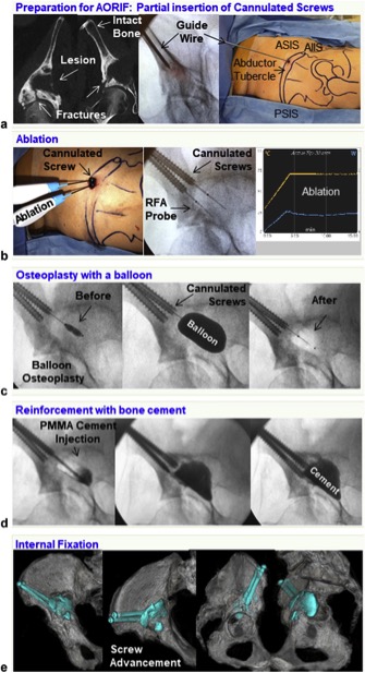
Figure 1. Illustrated details of the AORIF procedure.
Summary
Image guided ablation, osteoplasty, reinforcement, and internal fixation (AORIF) is a technique for treating and stabilizing osteolytic bone metastases. The procedure incorporates radiofrequency ablation for cancer treatment with PMMA cement and internal screw fixation for stabilization. This minimally invasive percutaneous approach aims to provide therapy, pain relief, and stability, while avoiding the risk associated with radiation and open surgery.
This study included 23 patients with 26 consecutive symptomatic osteolytic metastases who were treated with AORIF. Three patients had more than one treatment location. Lesions were located in the pelvis, proximal femur, proximal tibia, and calcaneus. AORIF was performed using cone beam CT guidance for 21 lesions and fluoroscopic guidance for 6 lesions. Post procedural pain and function, follow up imaging, and complications were evaluated.
No complications were described with the initial wire placement, screw placement, or ablation. Two balloons ruptured, which were retrieved without further complication. There was one instance of PMMA extravasation without further related complication. No intra or post procedural blood transfusion was required. No infection, wound complication, fracture or hardware complication was reported at 30 day follow up. No patient required a secondary or revision procedure during the follow-up period (1-18 months). All patients reported improved pain and function at 2 weeks post procedure.
Commentary
The authors evaluated 23 patients treated with AORIF for symptomatic lytic bone metastases. The results are impressive with all subjects experiencing improved pain and function, as well as zero postoperative complications, and no required secondary procedures during the follow-up period. While the cases of intraoperative balloon rupture and PMMA extravasation were inconsequential, they raise awareness for potential adverse events of the AORIF procedure. The study is limited by the small cohort, single center, and lack of control arm for comparison. Despite the limitations, the study demonstrates remarkable outcomes. The study highlights various benefits of AORIF including a spectrum of different anatomic applications and approaches. AORIF offers a low risk, minimally invasive surgical alternative for the management of lytic bone metastases. This study opens the door for future studies to further validate the AORIF procedure.
Post author
Maxwell R. Cretcher, DO
Resident Physician, Integrated Interventional Radiology
Dotter Department of Interventional Radiology
Oregon Health & Science University
No comments:
Post a Comment
Note: Only a member of this blog may post a comment.