MR Imaging-Guided Transurethral Ultrasound Ablation of Localized Prostate Cancer: Preliminary Experience from a Single Center in a Prospective, Multi-Center, Single-Arm Clinical Trial
Clinical question
Is magnetic resonance imaging (MRI)-guided transurethral ultrasound ablation (TULSA) of the prostate, with treatment parameters enabling ablation of almost the entire prostate (prior phase I trial with mandated ablation covering less than 90% of the prostate volume), safe and efficacious for the treatment of low- and intermediate-risk localized prostate cancer?
Take-away point
Yes, whole-gland MRI-guided TULSA of the prostate is safe and efficacious without significant quality of life impairment based on this preliminary trial with a 12-month follow-up.
Reference
MR Imaging-Guided Transurethral Ultrasound Ablation of Localized Prostate Cancer: Preliminary Experience from a Single Center in a Prospective, Multi-Center, Single-Arm Clinical Trial. Sundaram KM, Staruch R, Burtnyk M, Lane JS, Penson DF, Arora SS. Journal of Journal of Vascular and Interventional Radiology (JVIR), Volume 31, Issue 5, 740-746.
Click here for abstract
Study design
Single arm, retrospective, single-center study of 9 male patients with organ-confined low- and intermediate-risk disease who underwent whole-gland MRI-guided TULSA with a follow-up period of 12 months including safety, efficacy, and quality of life metrics.
Funding source
This trial was sponsored by Profound Medical (Mississauga, Ontario, Canada).
Setting
Academic hospital, Vanderbilt University Medical Center, Nashville, Tennessee.
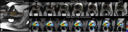
Figure 2. MRI-guided TULSA treatment workflow. (a) Sagittal T2-weighted planning images guide positioning of the transurethral ultrasound applicator and endorectal cooling device. (b) Axial T2-weighted images are used by the physician to contour a targeted prostate boundary (yellow line) for each transducer element. (c) Axial real-time MR thermometry images enable monitoring of the directional high-intensity ultrasound heating pattern and are used by the software to dynamically adjust treatment parameters for precise ablation to the prescribed boundaries.
Summary
Conventional treatments for localized prostate cancer have significant negative impacts on patients’ quality of life. MRI-guided TULSA of the prostate, a minimally invasive treatment modality, was previously demonstrated to be safe and feasible for conformal prostate ablation in a phase I trial. Given the nature of a feasibility study, a conservative approach requiring less than 90% coverage of the prostate volume was mandated. Safety and efficacy assessment of whole-gland TULSA, which is appropriate for multifocal and bilateral disease, was yet unavailable in the literature.
The authors performed a single-arm perspective single-center study of 9 male patients with localized, no greater than T2b, biopsy-confirmed adenocarcinoma of the prostate who underwent MRI-guided TULSA of the prostate. Additional important inclusion criteria included requirements on Gleason score, PSA, and prostate volume. Main exclusion criteria included tumor proximity to prostatic urethra or sphincter plane in the apex, prior treatments, significant prostatic cysts or calcifications, interests in future fertility, active infection, inflammatory conditions affecting the rectum, and severe neurogenic bladder or untreated bladder stones. Primary outcomes including incidence of treatment-emergent adverse events and PSA nadir as well as secondary outcomes including imaging appearance, 10-core biopsy, prostate volume, and quality of life assessment were collected at 12 months after the procedure.
All 9 patients underwent whole-gland TULSA with a planned sub-lethal thermal dose delivery to only 0.3% of the prostate volume. Procedure details were included in the published manuscript and the online supplemental material. Main pearls included 1) careful selection of patients (prostate volume, prostatic cysts, and calcifications assessment); 2) suprapubic catheter placement; 3) cooling device for protection of periurethral tissue and rectal wall, part of the TULSA-PRO system; 4) rectal air management; 5) meticulous positioning to maintain a 4-mm safety margin to the internal sphincter plane at the prostate apex; 6) repeated treatment to increase thermal dose (desiring at least 240 equivalent minutes at 43 degrees Celsius, or CEM43); 7) immediate post-treatment acute nonperfused volume assessment.
Median PSA reduction of 95.4% was observed. 1 patient with unexpectedly more extensive calcifications failed to reach the primary endpoint of more than 75% PSA reduction. There was few grade 2 adverse events and no grade 3 events. 12-month MRI showed 1 patient with highly suspicious lesion and 1 patient with mildly suspicious imaging findings. Biopsy showed histologic benefit in all 9 patients. All potent men remained potent without postprocedural changes in erectile firmness. Acute nonperfused volume was found to be predictive of 12-month PSA and non-small prostate (more than 18 cc) volume reduction.
Commentary
The authors in this paper presented their preliminary experience on the safety and efficacy of whole-gland TULSA in the treatment of low- and intermediate-risk localized prostate cancer. The trial was well-designed and data presentation well-constructed. Procedural details, especially the salient pearls and pitfalls, will be important for practice establishment and improvement. Results including minimal damage to surrounding tissue with proper techniques and good clinical outcome at 12-month follow-up by imaging, biopsy, labs, and patient self-reporting metrics were promising. Whole-gland TULSA may indeed be a good alternative treatment pathway between whole gland surgery/radiation and active surveillance. We eagerly anticipate the complete 12-month outcomes of the TULSA pivotal trial. Assessment on the safety and efficacy of focal TULSA ablative therapy should be conducted in a scientifically rigorous manner, as it was done by the authors of this study.
Post AuthorNingcheng (Peter) Li, MD, MS
Integrated Interventional Radiology Resident, PGY-3
Department of Interventional Radiology
Oregon Health and Science University, Dotter Interventional Institute
@NingchengLi
No comments:
Post a Comment
Note: Only a member of this blog may post a comment.