Monday, October 7, 2019

Identification of Parameters Influencing the Vascular Structure Displacement in Fusion Imaging during Endovascular Aneurysm Repair


Clinical question
Can we safely perform a zero-contrast procedure relying solely on fusion imaging? What contributes to renal ostia displacement by the insertion of a stiff system during endovascular aneurysm repair (EVAR) of abdominal aortic aneurysm (AAA)?

Take away point
Sharper angulation between aneurysm neck and sac results in decreased fusion imaging accuracy and precludes EVAR deployment without contrast validation.

Reference
Identification of Parameters Influencing the Vascular Structure Displacement in Fusion Imaging during Endovascular Aneurysm Repair. Lalys F. et al. Journal of Vascular and Interventional Radiology (JVIR), Volume 30, Issue 9, 1386-1392.

Click here for abstract

Study design
Single arm, prospective, cohort study of 50 patients undergoing EVAR.

Funding Source
No reported funding

Setting
Academic hospital, University Rennes, France.
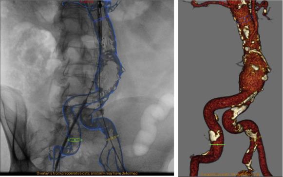


Figure 1
EndoNaut station main screen visualization during an EVAR procedure, with the fusion overlaid on the fluoroscopic image on the left and the corresponding 3D visualization on the right.

Summary

Fusion imaging technology has been shown to decrease contrast usage and radiation dose in both standard and complex endovascular aneurysm repair (EVAR) of abdominal aorta aneurysm (AAA). However, its relevance is limited by deformation of vascular structures after insertion of stiff devices during the procedure. The authors performed a prospective study of 50 patients undergoing EVAR to quantify the displacement of the lowest renal artery ostium and to identify the contributing anatomical parameters.

Patients included in this study were adults undergoing EVAR, who were not open-repair candidates, had AAA diameter greater than 50 mm, or had AAA growth rate greater than 10 mm per year. Patients were excluded if a fenestrated or branched endograft was used, or if the repair was performed for ruptured AAA.

EndoNaut station was used for fusion workflow. Digital subtraction angiogram performed intra-procedurally after insertion of stiff devices but prior to EVAR deployment was compared with the fusion roadmap generated pre-operatively using CT angiography. A large number of anatomical parameters were measured, including tortuosity index, length, diameter, angulation, calcification, thrombus, and distance to spine of the relevant vascular segments. Sharpest angulation, as defined by Van Keulen et al, was also calculated.

Mean displacement of the lowest renal artery ostium was 4.1 ± 2.4 mm (range, 0–11.7 mm), with a left/right displacement of 1.6 ± 1.7 mm (range, 0–6.9 mm) and a craniocaudal displacement of 3.5 ± 2.4 mm (range, 0–11.3 mm). Sharpest angulation between the aneurysmal neck and sac contributed significantly to the accuracy of fusion imaging (p = 0.00001).


Commentary

The authors in this study quantified the degree of renal artery ostia displacement after insertion of stiff devices during EVAR. They further identified the sharpest angulation between the aneurysmal neck and sac as a significant contributor in fusion imaging accuracy. The findings support the need for better fusion imaging technology with semi-automatic intra-procedural adjustments, especially in EVAR cases with significant angulation between the aneurysmal neck and sac. The paper also underlines the importance of small-amount contrast injection prior to EVAR deployment for confirmation of major vascular ostia. Future fusion imaging technology with more adaptive intra-procedural capability, potentially with the assistance of artificial intelligence, should be validated in a similar manner with prospective design and anatomical analysis before we proceed to attempt a zero-contrast EVAR deployment.

Post Author
Ningcheng (Peter) Li, MD, MS
Integrated Interventional Radiology Resident, PGY-3
Department of Interventional Radiology
Oregon Health and Science University, Dotter Interventional Institute
@NingchengLi

Friday, October 4, 2019

Safety and Efficacy of Acute Pulmonary Embolism Treated via Large-Bore Aspiration Mechanical Thrombectomy Using the Inari FlowTriever Device


Clinical question
Is it safe and effective to use the Inari FlowTriever Device in treating acute pulmonary embolism?

Take away point
Large-bore aspiration mechanical thrombectomy is safe with a high technical (100%) and clinical success (88%) rates.

Reference
Wible, Grant C. et al. Safety and Efficacy of Acute Pulmonary Embolism Treated via Large-Bore Aspiration Mechanical Thrombectomy Using the Inari FlowTriever Device. Journal of Vascular and Interventional Radiology, Volume 30, Issue 9, 1370-75

Click here for abstract

Study design
Single center, retrospective, cohort study of 46 patients between March 2018 – March 2019

Funding
One of the authors is a paid consultant for Inari and another receives research support from Inari.

Setting
St. Luke’s Hospital and University of Missouri – Kanas City school of medicine, Kansas City, MO, USA.


Figure 2
CT angiogram of a 68-year-old female with saddle pulmonary embolism (a). Right and left pulmonary angiogram demonstrating large filling defects corresponding to patients know PE (b,c). Post aspiration mechanical thrombectomy angiogram demonstrating significant improvement in pulmonary artery flow (d,e). Back table photographs of aspirated clot using the large-bore Inari device (f).


Summary


The authors performed a retrospective analysis of the Inari FlowTriever device at their institution when treating massive and intermediate-high risk sub-massive pulmonary embolism. They evaluated the technical success and clinical success of the device in the first year of use at their institution. Technical success involved appropriate device positioning and clinical success was defined by angiographic removal of central clot and change in pulmonary artery pressures before and after clot retrieval. Adverse outcomes were defined by the SIR guidelines of endovascular treatment of PE.

Patients included in this study were referred to IR for massive or intermediate-high risk sub-massive PE. No patients were excluded in the timeframe observed and 2 procedure-related adverse outcomes were reported.

All patients had aspiration mechanical thrombectomy with the Inari FlowTriever device with a 100% technical success rate. Pre and post-aspiration mechanical thrombectomy pressures were obtained, except in 4 cases, where the authors experienced failure of the pressure transducer. A 22F sheath was advanced into the suprarenal IVC via the common femoral vein and the 20F Inari aspiration guide catheter (AGC) was advanced over an 0.035 Amplatz wire into the main or lobar pulmonary artery. Aspiration thrombectomy was performed with the assistance of the self-expanding Nitinol discs to facilitate mechanical clot engagement prior to aspiration. This process was repeated several times in multiple pulmonary arteries until angiographic resolution of central clot was achieved.

There were 2 major procedure-related complications. One with intra-procedural hemoptysis requiring in intubation and another with a hematocrit drop of 15%. Both patients recovered and were discharged from the hospital. 2 patients died in the 30 day post-operative period, which the authors attributed to events prior to the procedure – metastatic pancreatic cancer and severe anoxic brain injury.

There was 100% technical success as defined by the authors. Aspiration related intra-procedure blood loss averaged 280ml with a maximum of 520ml resulting in an average hematocrit drop of 4.6% +/- 2.8% per case. Intra-procedural PA pressure decrease was achieved in 37 patients (88%). Average mean PA pressure improved from 33.9 +/- 8.9 mm Hg before to 27.0 +/- 9 mm Hg after, with a decrease of 6.9 mm Hg (P < 0.0001 and CI 5.0-8.8). All patients survived to hospital discharge.

Commentary


The authors evaluated the safety and efficacy of the Inari FlowTriever device at their institution over a one-year period. The findings of this study demonstrate the Inari device can be a treatment alternative in patients who are unable to receive systemic or local TPA in the setting of trauma, recent major surgery or other contraindication. This study only looked at a single centers experience with the device, which is useful in evaluating its success in the other interventional radiology departments. Establishing safety is a promising first step in assessing a new treatment paradigm, though comparison with systemic and local lytic and aspiration treatments is still needed.

Post Author
Hasnain Hasham, MD
Interventional Radiology Fellow
Dotter Interventional Institute
Oregon Health and Science University, Portland, OR
@irhashamz

Monday, September 30, 2019

Comparison of Imaging Changes and Pain Responses in Patients with Intra- or Extraosseous Bone Metastases Treated Palliatively with Magnetic Resonance- Guided High-Intensity–Focused Ultrasound


Clinical question
Does pain response differ in patients with intraosseous versus extraosseous bone metastases after ablation with MR guided High-Intensity-Focused Ultrasound (HIFU)? Can follow up MR predict treatment response?

Take away point
Patients with intraosseous tumors had significant improvement in pain from baseline after ablation with MR guided HIFU while to those with extraosseous tumors did not. Follow up with MR after ablation with HIFU does not predict efficacy of treatment.

Reference
Giles SL, Brown MRD, Rivens I, et al. Comparison of Imaging Changes and Pain Responses in Patients with Intra- or Extraosseous Bone Metastases Treated Palliatively with Magnetic Resonance-Guided High-Intensity-Focused Ultrasound. J Vasc Interv Radiol JVIR. 2019;30(9):1351-1360.e1. doi:10.1016/j.jvir.2019.02.019

Click here for abstract

Study design
Single arm, prospective study of 21 patients with dominant, painful bone metastasis (9 with intraosseous disease and 12 with extraosseous cortical breach) treated with MR guided HIFU.

Funding Source
Sponsor: Institute of Cancer Research, United Kingdom
Collaborators: Phillips Medical Systems, Cancer Research UK

Setting
Multicenter Study


Figure 4
Several scoring systems were utilized by the authors to evaluate post-procedure pain reduction. In this figure, the results of case report forms (CRFs) are shown for treated tumor for (a) 9 patients in the intraosseous group and (b) 12 patients in the extraosseous group. At days 30, 60, and 90 after treatment, scores were significantly lower than those in pretreatment (Pre Tx) for the intraosseous group but not for the extraosseous group.

Summary

High intensity focused ultrasound (HIFU) has shown promising results for palliative treatment of pain from bone metastases potentially related to thermal denervation of periosteum. The authors performed a prospective study of 21 patients with either interosseous or extraosseous painful bone metastasis who were then treated with MR guided HIFU. Gadolinium enhanced T1W images were obtained at the completion of treatment and at 30, 60 and 90 day follow up. Patient pain was assessed using various scoring systems at the time of the procedure and at follow up in addition to recorded analgesic use, which was compared before and after ablation. Patients were categorized as responders and non-responders in regard to post procedure alleviation of pain.

A difference from baseline pain was only statistically significant for patients with interosseous tumor on follow up. While 67% of patients with interosseous tumors were classified as responders at 30 days, only 33% of patients in the extraosseous group responded.

While imaging results of the ablated tumor showed nonenhancement in the majority of interosseous tumors, post procedure enhancement patterns of ablated extraosseous tumors varied. There was no difference in follow up imaging enhancement characteristics for patients who were classified as responders or non-responders.

Commentary

The authors in this study evaluated 21 patients with painful bone metastasis after treatment with MR guided High Intensity Focused Ultrasound. While the findings are encouraging for patients with intraosseous disease where the bone cortex remains undisrupted with significant improvement in pain for 67% of their patients at 30 days, results for extraosseous extension of disease were less promising. The authors alluded to the fact that patients with extraosseous disease may have more advanced disease at the time of treatment and more studies evaluating ablative thermal dose per tumor volume in these patients are required. While follow up MR may be beneficial for monitoring disease progression, preliminary results from this study showed that MR could not predict efficacy of treatment. As noted by the authors, in the future, this finding may point to fewer burdensome MRs obtained after ablation.

Post Author
Teodora Bochnakova MD
Assistant Professor
Department of Interventional Radiology
Oregon Health and Science University, Portland, OR
@T_bochnakova